Difference between revisions of "Kimura disease"
Jump to navigation
Jump to search
m |
|||
| (4 intermediate revisions by 2 users not shown) | |||
| Line 1: | Line 1: | ||
{{ Infobox diagnosis | {{ Infobox diagnosis | ||
| Name = {{PAGENAME}} | | Name = {{PAGENAME}} | ||
| Image = Kimura_disease_-_very_high_mag.jpg | | Image = Kimura_disease_-_very_high_mag.jpg | ||
| Width = | | Width = | ||
| Caption = Kimura disease. [[H&E stain]]. | | Caption = Kimura disease. [[H&E stain]]. | ||
| Micro = eosinophils and thick walled [[blood vessel]]s with | | Micro = eosinophils and thick walled [[blood vessel]]s with [[hobnail]]ed endothelial cells | ||
| Subtypes = | | Subtypes = | ||
| LMDDx = [[angiolymphoid hyperplasia with eosinophilia]], [[drug reaction]], infection (parasitic), [[lymphoma]] | | LMDDx = [[angiolymphoid hyperplasia with eosinophilia]], [[drug reaction]], infection (parasitic), [[lymphoma]], [[LCH]] | ||
| Stains = | | Stains = | ||
| IHC = | | IHC = | ||
| Line 15: | Line 15: | ||
| Grossing = | | Grossing = | ||
| Site = [[lymph node]], head and neck | | Site = [[lymph node]], head and neck | ||
| Assdx = | |||
| Syndromes = | |||
| Clinicalhx = | |||
| Signs = | | Signs = | ||
| Symptoms = | | Symptoms = | ||
| Line 44: | Line 47: | ||
Features:<ref name=Ref_ILNP190>{{Ref ILNP|190}}</ref> | Features:<ref name=Ref_ILNP190>{{Ref ILNP|190}}</ref> | ||
*Angiolymphoid proliferation. | *Angiolymphoid proliferation. | ||
**Thick walled blood vessels with (plump) hobnail endothelial cells.<ref>URL: [http://emedicine.medscape.com/article/1098777-diagnosis http://emedicine.medscape.com/article/1098777-diagnosis]. Accessed on: 8 August 2010.</ref> | **Thick walled blood vessels with (plump) [[hobnail]] endothelial cells.<ref>URL: [http://emedicine.medscape.com/article/1098777-diagnosis http://emedicine.medscape.com/article/1098777-diagnosis]. Accessed on: 8 August 2010.</ref> | ||
*Eosinophils - abundant - '''key feature'''. | *Eosinophils - abundant - '''key feature'''. | ||
| Line 62: | Line 65: | ||
Image:Kimura_disease_-_intermed_mag.jpg | Kimura disease - intermed. mag. (WC) | Image:Kimura_disease_-_intermed_mag.jpg | Kimura disease - intermed. mag. (WC) | ||
</gallery> | </gallery> | ||
==IHC== | ==IHC== | ||
*Used to rule-out a clonal population, i.e. [[lymphoma]]. | *Used to rule-out a clonal population, i.e. [[lymphoma]]. | ||
Latest revision as of 03:01, 31 October 2015
| Kimura disease | |
|---|---|
| Diagnosis in short | |
 Kimura disease. H&E stain. | |
|
| |
| LM | eosinophils and thick walled blood vessels with hobnailed endothelial cells |
| LM DDx | angiolymphoid hyperplasia with eosinophilia, drug reaction, infection (parasitic), lymphoma, LCH |
| Site | lymph node, head and neck |
|
| |
| Prevalence | extremely rare |
| Blood work | eosinophilia |
Kimura disease is a rare disease with abundant eosinophils. It may show-up in a lymph node specimen. It is similar to angiolymphoid hyperplasia with eosinophilia.[1]
General
- AKA eosinophilic lymphogranuloma, Kimura disease.
- Chronic inflammatory disorder - suspected to be infectious.
Clinical:
- Usually neck, periauricular.
- Peripheral blood eosinophilia.
- Increased blood IgE.
Epidemiology
- Males > females.
- Young.
- Asian.
Microscopic
Features:[2]
- Angiolymphoid proliferation.
- Eosinophils - abundant - key feature.
DDx:
- Drug reaction.
- Parasitic infection.
- Angiolymphoid hyperplasia with eosinophilia.
Notes:
- In a lymph node... it may be signed-out as reactive lymphadenitis with follicular hyperplasia and prominent eosinophils, see comment.
- Abundant eosinophils: consider Langerhans cell histiocytosis.
Images
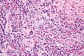
Kimura disease - very high mag. (WC)
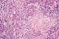
Kimura disease - high mag. (WC)
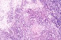
Kimura disease - intermed. mag. (WC)


IHC
- Used to rule-out a clonal population, i.e. lymphoma.
See also
References
- ↑ URL: http://emedicine.medscape.com/article/1082603-overview. Accessed on: 14 January 2012.
- ↑ Ioachim, Harry L; Medeiros, L. Jeffrey (2008). Ioachim's Lymph Node Pathology (4th ed.). Lippincott Williams & Wilkins. pp. 190. ISBN 978-0781775960.
- ↑ URL: http://emedicine.medscape.com/article/1098777-diagnosis. Accessed on: 8 August 2010.