Kaposi sarcoma
Jump to navigation
Jump to search
| Kaposi sarcoma | |
|---|---|
| Diagnosis in short | |
 Kaposi sarcoma. H&E stain. | |
|
| |
| LM | vascular lesion (abundant RBCs), +/-"promontory sign", +/-spindle cells with minimal nuclear atypia, RBC extravasation, +/-intracytoplasmic hyaline globules, +/-hemosiderin deposits, +/-plasma cells |
| Subtypes | classic, endemic, immunosuppression-associated or transplant-associated, AIDS-associated |
| LM DDx | angiosarcoma, Masson's hemangioma (intravascular papillary endothelial hyperplasia), benign lymphangioendothelioma |
| IHC | HHV-8 +ve, CD31 +ve ,CD34 +ve |
| Gross | red or brown patches, plaques or nodules |
| Site | skin, others |
|
| |
| Associated Dx | +/-HIV infection or immunoincompentence |
| Prevalence | uncommon |
| Prognosis | dependent on subtype |
Kaposi sarcoma, abbreviated KS, is an uncommon vascular tumour that is often associated with HIV/AIDS.
General
- Caused by Human herpesvirus-8 (HHV-8).
- In the North American context, it is often associated with immunodeficiency, e.g. HIV/AIDS.
Interesting note:
- It has been said that KS is not really a sarcoma.[1]
Stages
It is seen in different stages:[2][3]
- Patch stage.
- Plaque stage.
- Nodular stage.
- Exophytic stage.
- Infiltrative stage.
- Lymphadenopathic stage.
Note:
- The first three are the classic ones.
Type or form
Classically divided into four types:[4][5][6]
- Classic = old men Mediterranean or Ashkenazi Jew.
- Endemic = African infants and young males.
- Immunosuppression-associated or transplant-associated - iatrogenic.
- AIDS-associated.
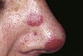
Gross
- Skin: red or brown patches, plaques or nodules.
KS. (WC)

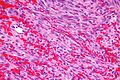
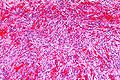
Microscopic
Features:[7]
- Vascular lesion (abundant RBCs) with:
- +/-"Promontory sign" - small vessel protruding into an abnormal vascular space.[8]
- Not pathognomonic for KS.[9]
- +/-Spindle cells with minimal nuclear atypia.
- RBC extravasation - very useful - important feature.[10]
- +/-"Promontory sign" - small vessel protruding into an abnormal vascular space.[8]
- +/-Intracytoplasmic hyaline globules - uncommon - one usu. needs to search for 'em.[11]
- Pale pink globs (that are paler than RBCs) - important feature.
- +/-Hemosiderin deposits.
- +/-Plasma cells.[12]
Notes:
- Hyaline globules have a DDx (hepatocellular carcinoma, lung adenocarcinoma, chondrosarcomas + others).[11]
DDx:
- Angiosarcoma - have many mitoses, nuclear atypia, RBC extravasation not common.
- Masson's hemangioma - AKA intravascular papillary endothelial hyperplasia.
- Benign lymphangioendothelioma.[13]
- Histologically very similar.[14]
- Cavernous hemangioma.[15]
Images
Kaposi sacoma - high mag. (WC)
Kaposi sarcoma - intermed. mag. (WC)

www:
Stains
- PAS +ve -- hyaline globules.
IHC
Features:[15]
See also
References
- ↑ Pérez, A.; Sánchez, JL.; Almodóvar, PI. (Oct 2003). "Kaposi's sarcoma is not a neoplasm let alone a sarcoma.". Int J Dermatol 42 (10): 844-5. PMID 14521707.
- ↑ URL: http://www.histopathology-india.net/KS.htm. Accessed on: 31 January 2010.
- ↑ URL: http://emedicine.medscape.com/article/1083998-clinical#a0217. Accessed on: 17 November 2011.
- ↑ Szajerka, T.; Jablecki, J.. "Kaposi's sarcoma revisited.". AIDS Rev 9 (4): 230-6. PMID 18219366.
- ↑ Morand, JJ.; Lightburn, E.; Simon, F.; Patte, JH. (Apr 2007). "[Update on Kaposi's sarcoma].". Med Trop (Mars) 67 (2): 123-30. PMID 17691428.
- ↑ Antman, K.; Chang, Y. (Apr 2000). "Kaposi's sarcoma.". N Engl J Med 342 (14): 1027-38. doi:10.1056/NEJM200004063421407. PMID 10749966.
- ↑ Klatt, Edward C. (2006). Robbins and Cotran Atlas of Pathology (1st ed.). Saunders. pp. 23. ISBN 978-1416002741.
- ↑ Lazova R, McNiff JM, Glusac EJ, Godic A (April 2009). "Promontory sign--present in patch and plaque stage of angiosarcoma!". Am J Dermatopathol 31 (2): 132–6. doi:10.1097/DAD.0b013e3181951045. PMID 19318797.
- ↑ Fernandez-Flores A, Rodriguez R (June 2010). "Promontory Sign in a Reactive Benign Vascular Proliferation". Am J Dermatopathol. doi:10.1097/DAD.0b013e3181cf0ae5. PMID 20577080.
- ↑ Kato, H.; Hamada, T.; Tsuji, T.; Baba, T.; Seki, J.; Kobayashi, Y. (Jul 1990). "Kaposi's sarcoma: a light and electron microscopic study.". J Dermatol 17 (7): 414-22. PMID 2229644.
- ↑ 11.0 11.1 del Rosario AD, Bui HX, Singh J, Ginsburg R, Ross JS (December 1994). "Intracytoplasmic eosinophilic hyaline globules in cartilaginous neoplasms: a surgical, pathological, ultrastructural, and electron probe x-ray microanalytic study". Hum. Pathol. 25 (12): 1283–9. PMID 7528163.
- ↑ Douglas, JL.; Gustin, JK.; Dezube, B.; Pantanowitz, JL.; Moses, AV. (Sep 2007). "Kaposi's sarcoma: a model of both malignancy and chronic inflammation.". Panminerva Med 49 (3): 119-38. PMID 17912148.
- ↑ Guillou, L.; Fletcher, CD. (Aug 2000). "Benign lymphangioendothelioma (acquired progressive lymphangioma): a lesion not to be confused with well-differentiated angiosarcoma and patch stage Kaposi's sarcoma: clinicopathologic analysis of a series.". Am J Surg Pathol 24 (8): 1047-57. PMID 10935645.
- ↑ URL: http://path.upmc.edu/cases/case134/dx.html. Accessed on: 5 January 2012.
- ↑ 15.0 15.1 Onak Kandemir N, Barut F, Doğan Gün B, Solak Tekin N, Hallaç Keser S, Oğuz Özdamar S (2013). "Cavernous hemangioma-like kaposi sarcoma: histomorphologic features and differential diagnosis". Case Rep Med 2013: 959812. doi:10.1155/2013/959812. PMC 3800618. PMID 24187557. https://www.ncbi.nlm.nih.gov/pmc/articles/PMC3800618/.
- ↑ Wada DA, Perkins SL, Tripp S, Coffin CM, Florell SR (February 2007). "Human herpesvirus 8 and iron staining are useful in differentiating Kaposi sarcoma from interstitial granuloma annulare". Am. J. Clin. Pathol. 127 (2): 263–70. doi:10.1309/GMH9CENH4909AWVB. PMID 17210517.