Difference between revisions of "Diffuse astrocytoma"
Jensflorian (talk | contribs) (update) |
Jensflorian (talk | contribs) (WHO 2016 update) |
||
| Line 5: | Line 5: | ||
* Usually shows progression to [[glioblastoma]] sooner or later. | * Usually shows progression to [[glioblastoma]] sooner or later. | ||
WHO 2016 categorization combines morphology and genetics into following groups:<ref>{{Cite journal | last1 = Louis | first1 = DN. | last2 = Perry | first2 = A. | last3 = Reifenberger | first3 = G. | last4 = von Deimling | first4 = A. | last5 = Figarella-Branger | first5 = D. | last6 = Cavenee | first6 = WK. | last7 = Ohgaki | first7 = H. | last8 = Wiestler | first8 = OD. | last9 = Kleihues | first9 = P. | title = The 2016 World Health Organization Classification of Tumors of the Central Nervous System: a summary. | journal = Acta Neuropathol | volume = 131 | issue = 6 | pages = 803-20 | month = Jun | year = 2016 | doi = 10.1007/s00401-016-1545-1 | PMID = 27157931 }}</ref> | |||
*Diffuse astrocytoma ICD-O: 9400/3 | *Diffuse astrocytoma, IDH-mutant ICD-O: 9400/3 - most frequent. | ||
** | **Gemistocytic astrocytoma, IDH-mutant ICD-O:9411/3 | ||
* | *Diffuse astrocytoma, IDH-wildtype ICD-O: 9400/3 | ||
* | *Diffuse astrocytoma,NOS ICD-O: 9400/3 - genetic data missing. | ||
Note: This subtyping is no longer in use | |||
''Note:'' Older terminologies included Fibrillary astrocytoma (ICD-O: 9420/3) and Protoplasmatic astrocytoma (ICD-O:9410/3)<ref name=WHOCNS>{{Ref WHOCNS|25}}</ref> This subtyping is no longer in use. These tumors are now classified according their IDH mutation status. | |||
==Radiology/Clinic== | ==Radiology/Clinic== | ||
| Line 44: | Line 45: | ||
File:Astrocytoma whoII HE.jpg | Astrocytoma, fibrillary type (WC/jensflorian) | File:Astrocytoma whoII HE.jpg | Astrocytoma, fibrillary type (WC/jensflorian) | ||
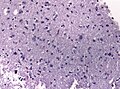
File:Neuropathology case II 02.jpg | Astrocytoma, protoplasmatic type (WC/jensflorian) | File:Neuropathology case II 02.jpg | Astrocytoma, protoplasmatic type (WC/jensflorian) | ||
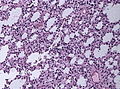
File:Gemistocytic astrocytoma.jpg | Gemistocytic astrocytoma (WC/jensflorian) | |||
</gallery> | </gallery> | ||
| Line 55: | Line 57: | ||
*MIB-1: 0-5% (mean: 2%). | *MIB-1: 0-5% (mean: 2%). | ||
*[[IDH-1]] (R132H)+ve in 60-70%. | *[[IDH-1]] (R132H)+ve in 60-70%. | ||
*[[ATRX]] loss in 70%. | **'Note:'' This antibody does not detect other rare IDH1/2 mutations. | ||
*[[ATRX]] nuclear loss in 70%. | |||
<gallery> | <gallery> | ||
| Line 63: | Line 66: | ||
==Molecular== | ==Molecular== | ||
*IDH1 R132- or IDH2 R172-point mutations classify the tumors as Diffuse astrocytoma, IDH-mutant. | |||
*Absence of LOH 1p/19q. | *Absence of LOH 1p/19q. | ||
*Tp53 mutations in approx. 60% (80-90% in gemistocytic, 50% in fibrillary types). | *Tp53 mutations in approx. 60% (80-90% in gemistocytic, 50% in fibrillary types). | ||
| Line 70: | Line 74: | ||
*Reactive astrocytosis. | *Reactive astrocytosis. | ||
*Demyelinisation. | *Demyelinisation. | ||
*[[Anaplastic astrocytoma]] | *[[Anaplastic astrocytoma]] - increased mitotic activity. | ||
*[[Oligoastrocytoma]] | *[[Oligoastrocytoma]], NOS - esp. when genetic data on IDH and LOH 1p/19q are lacking. | ||
*[[Oligodendroglioma]] - esp. protoplasmatic forms. | *[[Oligodendroglioma]] - esp. protoplasmatic forms. LOH 1p/19q testing required. | ||
*[[SEGA]] - esp. gemistocytic forms. | *[[SEGA]] - esp. gemistocytic forms. | ||
Revision as of 08:14, 19 May 2016
Diffuse astrocytoma (AKA: diffuse, low-grade astrocytoma) is a infiltrating astrocytoma occurring in the CNS white matter.
- Most common grade II WHO glioma in adults (peaks between 30-40 years).
- 10-15% of all astrocytomas.
- Usually shows progression to glioblastoma sooner or later.
WHO 2016 categorization combines morphology and genetics into following groups:[1]
- Diffuse astrocytoma, IDH-mutant ICD-O: 9400/3 - most frequent.
- Gemistocytic astrocytoma, IDH-mutant ICD-O:9411/3
- Diffuse astrocytoma, IDH-wildtype ICD-O: 9400/3
- Diffuse astrocytoma,NOS ICD-O: 9400/3 - genetic data missing.
Note: Older terminologies included Fibrillary astrocytoma (ICD-O: 9420/3) and Protoplasmatic astrocytoma (ICD-O:9410/3)[2] This subtyping is no longer in use. These tumors are now classified according their IDH mutation status.
Radiology/Clinic
- Mass effect.
- Seizures.
- Neurologic decifit.
- Usually not contrast-enhanching, T2 bright.
Macroscopy
- No clear demarcation from white matter
- May contain larger cysts
- No necrosis
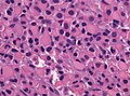
Histology
Features: [3]
- Cell density higher than normal brain.
- Mild to moderate nuclear pleomorphism.
- Monotony of atypical nuclei and irregular distribution indicates neoplasm.
- "naked nuclei" without recognizeable processes.
- No prominent nucleolus.
- Cytoplasm highly variable (even within the same tumour).
- In normal CNS the cytoplasm blends within the neuropil.
- Mitoses absent or very rare.
- Microcystic spaces of the background (none to extensive).
- No necrosis, no vascular proliferations.
- Except radiation necrosis.
- Lymphocytic cuffing (mostly in gemistocytic type)
- Abent to few rosenthal fibers.
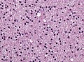
Diffuse astrocytoma, H&E (WC/jensflorian)
Diffuse astrocytoma, H&E (WC/jensflorian)
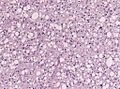
Astrocytoma, fibrillary type (WC/jensflorian)
Astrocytoma, protoplasmatic type (WC/jensflorian)
Gemistocytic astrocytoma (WC/jensflorian)


IHC
- GFAP+ve.
- MAP2+ve (especially in cell processes).
- Vimentin+ve (often perinuclear).
- S-100+ve.
- p53: Nuclear staining in 30% of the tumours (usually few cells).
- MIB-1: 0-5% (mean: 2%).
- IDH-1 (R132H)+ve in 60-70%.
- 'Note: This antibody does not detect other rare IDH1/2 mutations.
- ATRX nuclear loss in 70%.
GFAP in astrocytoma (WC/jensflorian)
ATRX loss in astrocytoma (WC/jensflorian)


Molecular
- IDH1 R132- or IDH2 R172-point mutations classify the tumors as Diffuse astrocytoma, IDH-mutant.
- Absence of LOH 1p/19q.
- Tp53 mutations in approx. 60% (80-90% in gemistocytic, 50% in fibrillary types).
- MGMT promotor methylated in approx. 50%.
DDx
- Reactive astrocytosis.
- Demyelinisation.
- Anaplastic astrocytoma - increased mitotic activity.
- Oligoastrocytoma, NOS - esp. when genetic data on IDH and LOH 1p/19q are lacking.
- Oligodendroglioma - esp. protoplasmatic forms. LOH 1p/19q testing required.
- SEGA - esp. gemistocytic forms.
See also
- ↑ Louis, DN.; Perry, A.; Reifenberger, G.; von Deimling, A.; Figarella-Branger, D.; Cavenee, WK.; Ohgaki, H.; Wiestler, OD. et al. (Jun 2016). "The 2016 World Health Organization Classification of Tumors of the Central Nervous System: a summary.". Acta Neuropathol 131 (6): 803-20. doi:10.1007/s00401-016-1545-1. PMID 27157931.
- ↑ The International Agency for Research on Cancer (Editors: Louis, D.N.; Ohgaki, H.; Wiestler, O.D.; Cavenee, W.K.) (2007). Pathology and Genetics of Tumours of Tumors of the Central Nervous System (IARC WHO Classification of Tumours) (4th ed.). Lyon: World Health Organization. pp. 25. doi:10.1007/s00401-007-0243-4. ISBN 978-9283224303.
- ↑ Burger, P.C.; Scheithauer, B.W. (2007). Tumors of the Central Nervous System (Afip Atlas of Tumor Pathology) (4th ed.). Washington: American Registry of Pathology. pp. 34. ISBN 1933477016.