Difference between revisions of "Chromophobe renal cell carcinoma"
(→IHC) |
|||
| (35 intermediate revisions by the same user not shown) | |||
| Line 1: | Line 1: | ||
{{ Infobox diagnosis | {{ Infobox diagnosis | ||
| Name = {{PAGENAME}} | | Name = {{PAGENAME}} | ||
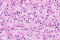
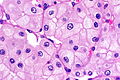
| Image = | | Image = Chromophobe renal cell carcinoma -- high mag.jpg | ||
| Width = | | Width = | ||
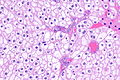
| Caption = | | Caption = Chromophobe renal cell carcinoma showing the characteristic perinuclear clearing and distinctive (plant-like) cellular borders. [[H&E stain]]. | ||
| Micro = pale cytoplasm | | Micro = pale/clear (or eosinophilic) cytoplasm with wisps of eosinophilic material, perinuclear clearing (a pale halo surrounds the nucleus), periphery of cell distinct (cell membrane is easy to discern -- plant cell-like) | ||
| Subtypes = classic, eosinophilic variant | | Subtypes = classic, eosinophilic variant | ||
| LMDDx = [[clear cell renal cell carcinoma]] (classic), [[renal oncocytoma]], [[clear cell renal cell carcinoma]] eosinophilic variant | | LMDDx = [[clear cell renal cell carcinoma]] (classic), [[renal oncocytoma]], [[clear cell renal cell carcinoma]] eosinophilic variant, [[renal hybrid oncocytic/chromophobe tumour]], other [[renal tumours with eosinophilic cytoplasm]] | ||
| Stains = [[Hale's colloidal iron]] +ve | | Stains = [[Hale's colloidal iron]] ([[Mueller-Mowry stain]]) +ve | ||
| IHC = CK7 +ve cell membrane, CD117 +ve, vimentin -ve | | IHC = CK7 +ve cell membrane, CD117 +ve, vimentin -ve | ||
| EM = | | EM = | ||
| Molecular = | | Molecular = | ||
| IF = | | IF = | ||
| Gross = | | Gross = grey-beige, lacks central scar | ||
| Grossing = | | Grossing = [[total nephrectomy for tumour grossing]], [[partial nephrectomy grossing]] | ||
| Staging = [[kidney cancer staging]] | |||
| Site = [[kidney]] - see [[renal tumours]] | | Site = [[kidney]] - see [[renal tumours]] | ||
| Assdx = | | Assdx = | ||
| Line 26: | Line 27: | ||
| Prognosis = | | Prognosis = | ||
| Other = | | Other = | ||
| ClinDDx = | | ClinDDx = other [[renal tumours]] | ||
| Tx = surgical resection | |||
}} | }} | ||
'''Chromophobe renal cell carcinoma''', abbreviated '''ChRCC''', is a relatively common form of [[renal cell carcinoma]]. | '''Chromophobe renal cell carcinoma''', abbreviated '''ChRCC''', is a relatively common form of [[renal cell carcinoma]]. | ||
| Line 32: | Line 34: | ||
==General== | ==General== | ||
*Least common of the common types of RCC ([[clear cell RCC]], [[papillary RCC]], [[chromophobe RCC]]). | *Least common of the common types of RCC ([[clear cell RCC]], [[papillary RCC]], [[chromophobe RCC]]). | ||
*''Fuhrman grading'' for this entity | *[[ISUP nucleolar grade|ISUP nucleolar grading]] (replaces ''Fuhrman grading'') not done for this entity, as it does not appear to have any predictive value.<ref name=pmid17527087>{{Cite journal | last1 = Delahunt | first1 = B. | last2 = Sika-Paotonu | first2 = D. | last3 = Bethwaite | first3 = PB. | last4 = McCredie | first4 = MR. | last5 = Martignoni | first5 = G. | last6 = Eble | first6 = JN. | last7 = Jordan | first7 = TW. | title = Fuhrman grading is not appropriate for chromophobe renal cell carcinoma. | journal = Am J Surg Pathol | volume = 31 | issue = 6 | pages = 957-60 | month = Jun | year = 2007 | doi = 10.1097/01.pas.0000249446.28713.53 | PMID = 17527087 }}</ref> | ||
*May be associated with [[Birt–Hogg–Dubé syndrome]].<ref name=Ref_WMSP290>{{Ref WMSP|290}}</ref> | *May be associated with [[Birt–Hogg–Dubé syndrome]].<ref name=Ref_WMSP290>{{Ref WMSP|290}}</ref> | ||
*Can be seen in the context of [[renal oncocytosis]].<ref name=pmid23018240>{{Cite journal | last1 = Kuroda | first1 = N. | last2 = Tanaka | first2 = A. | last3 = Ohe | first3 = C. | last4 = Mikami | first4 = S. | last5 = Nagashima | first5 = Y. | last6 = Sasaki | first6 = T. | last7 = Inoue | first7 = K. | last8 = Hes | first8 = O. | last9 = Michal | first9 = M. | title = Review of renal oncocytosis (multiple oncocytic lesions) with focus on clinical and pathobiological aspects. | journal = Histol Histopathol | volume = 27 | issue = 11 | pages = 1407-12 | month = Nov | year = 2012 | doi = | PMID = 23018240 }}</ref> | |||
===Subtypes=== | |||
There are two subtypes:<ref name=Ref_GUP293>{{Ref GUP|293}}</ref> | There are two subtypes:<ref name=Ref_GUP293>{{Ref GUP|293}}</ref> | ||
*Classic. | *Classic. | ||
| Line 40: | Line 44: | ||
==Gross== | ==Gross== | ||
* | *Grey-beige colour.<ref name=pmid12507296>{{Cite journal | last1 = Kuroda | first1 = N. | last2 = Toi | first2 = M. | last3 = Hiroi | first3 = M. | last4 = Enzan | first4 = H. | title = Review of chromophobe renal cell carcinoma with focus on clinical and pathobiological aspects. | journal = Histol Histopathol | volume = 18 | issue = 1 | pages = 165-71 | month = Jan | year = 2003 | doi = | PMID = 12507296 }}</ref> | ||
*Solitary. | *Solitary, usually.‡ | ||
*Well-circumscribed. | *Well-circumscribed. | ||
Image | Note: | ||
*‡ Approximately 3% are multifocal.<ref name=pmid22502873>{{Cite journal | last1 = Siracusano | first1 = S. | last2 = Novara | first2 = G. | last3 = Antonelli | first3 = A. | last4 = Artibani | first4 = W. | last5 = Bertini | first5 = R. | last6 = Carini | first6 = M. | last7 = Carmignani | first7 = G. | last8 = Ciciliato | first8 = S. | last9 = Cunico | first9 = SC. | title = Prognostic role of tumour multifocality in renal cell carcinoma. | journal = BJU Int | volume = 110 | issue = 11 Pt B | pages = E443-8 | month = Dec | year = 2012 | doi = 10.1111/j.1464-410X.2012.11121.x | PMID = 22502873 }}</ref> | |||
===Image=== | |||
*[http://www.flickr.com/photos/35441329@N05/4273199789/in/photostream/ Chromophobe RCC (flickr.com)]. | *[http://www.flickr.com/photos/35441329@N05/4273199789/in/photostream/ Chromophobe RCC (flickr.com)]. | ||
==Microscopic== | ==Microscopic== | ||
===Classic=== | ===Classic=== | ||
Features - classic type (3 P's | Features - classic type (3 P's memory device):<ref>{{Ref PBoD|1016-7}}</ref><ref name=Ref_GUP293>{{Ref GUP|293}}</ref> | ||
*Pale cytoplasm, with wisps of eosinophilic material; the cells are not completely clear, they have "cobwebs". | *Pale cytoplasm, with wisps of eosinophilic material; the cells are not completely clear, they have "cobwebs". | ||
*Perinuclear clearing, i.e. a pale halo surrounds the nucleus - '''key feature'''. | *Perinuclear clearing, i.e. a pale halo surrounds the nucleus - '''key feature'''. | ||
| Line 63: | Line 70: | ||
**Perinuclear clearing is ''not'' seen in clear cell RCC. | **Perinuclear clearing is ''not'' seen in clear cell RCC. | ||
**ChRCC has wisps in the cytoplasm. | **ChRCC has wisps in the cytoplasm. | ||
*Other [[clear cell tumours]]. | |||
*[[Benign clear cell clusters of the kidney]] - somewhat controversial. | |||
====Images==== | |||
<gallery> | |||
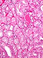
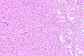
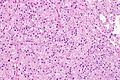
Image: Chromophobe renal cell carcinoma -- low mag.jpg | ChRCC - low mag. (WC/Nephron) | |||
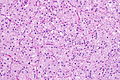
Image: Chromophobe renal cell carcinoma -- intermed mag.jpg | ChRCC - intermed. mag. (WC/Nephron) | |||
Image: Chromophobe renal cell carcinoma -- high mag.jpg | ChRCC - high mag. (WC/Nephron) | |||
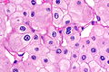
Image: Chromophobe renal cell carcinoma -- very high mag.jpg | ChRCC - very high mag. (WC/Nephron) | |||
Image: Chromophobe renal cell carcinoma - alt -- high mag.jpg | ChRCC - high mag. (WC/Nephron) | |||
Image: Chromophobe renal cell carcinoma - alt -- very high mag.jpg | ChRCC - very high mag. (WC/Nephron) | |||
</gallery> | |||
===Eosinophilic variant=== | ===Eosinophilic variant=== | ||
| Line 82: | Line 101: | ||
#*Perinuclear clearing is ''not'' seen in clear cell RCC. | #*Perinuclear clearing is ''not'' seen in clear cell RCC. | ||
#*ChRCC has wisps in the cytoplasm. | #*ChRCC has wisps in the cytoplasm. | ||
#[[Renal hybrid oncocytic/chromophobe tumour]]. | |||
#Other [[renal tumours with eosinophilic cytoplasm]]. | |||
====Images==== | |||
=====Case 1===== | |||
<gallery> | |||
Image:Chromophobe renal cell carcinoma, eosinophilic variant - high mag.jpg | Eosinophilic chromophobe RCC. (WC/Nephron) | |||
</gallery> | |||
=== | =====Case 2===== | ||
<gallery> | <gallery> | ||
Image: | Image: Eosinophilic variant of chromophobe renal cell carcinoma -- low mag.jpg | EVChRCC - low mag. | ||
</gallery> | Image: Eosinophilic variant of chromophobe renal cell carcinoma -- intermed mag.jpg | EVChRCC - intermed. mag. | ||
www | Image: Eosinophilic variant of chromophobe renal cell carcinoma - alt -- intermed mag.jpg | EVChRCC - intermed. mag. | ||
Image: Eosinophilic variant of chromophobe renal cell carcinoma -- high mag.jpg | EVChRCC - high mag. | |||
Image: Eosinophilic variant of chromophobe renal cell carcinoma -- very high mag.jpg | EVChRCC - very high mag. | |||
Image: Eosinophilic variant of chromophobe renal cell carcinoma - alt -- very high mag.jpg | EVChRCC - very high mag. | |||
</gallery> | |||
=====www===== | |||
*[http://path.upmc.edu/cases/case333.html Chromophobe RCC - several images (upmc.edu)]. | *[http://path.upmc.edu/cases/case333.html Chromophobe RCC - several images (upmc.edu)]. | ||
==Stains== | ==Stains== | ||
*[[Hale's colloidal iron]] +ve (blue granular cytoplasmic). | *[[Hale's colloidal iron]] +ve (blue granular cytoplasmic). | ||
**Oncocytoma -ve. | |||
Note: | |||
*This seems to be a difficult stain to get working. | |||
Images | ===Images=== | ||
*[http://www.nature.com/modpathol/journal/v18/n2/fig_tab/3800286f1.html ChRCC Hale's colloidal iron - several images (nature.com)]. | *[http://www.nature.com/modpathol/journal/v18/n2/fig_tab/3800286f1.html ChRCC Hale's colloidal iron - several images (nature.com)]. | ||
*[http://www.ultrapath.org/oldsite/cases99/sep99/images/figure-3.jpg ChRCC Hale's colloidal iron (ultrapath.org)].<ref>URL: [http://www.ultrapath.org/oldsite/cases99/sep99/cotm9-2.html http://www.ultrapath.org/oldsite/cases99/sep99/cotm9-2.html]. Accessed on: 9 October 2011.</ref> | *[http://www.ultrapath.org/oldsite/cases99/sep99/images/figure-3.jpg ChRCC Hale's colloidal iron (ultrapath.org)].<ref>URL: [http://www.ultrapath.org/oldsite/cases99/sep99/cotm9-2.html http://www.ultrapath.org/oldsite/cases99/sep99/cotm9-2.html]. Accessed on: 9 October 2011.</ref> | ||
| Line 99: | Line 134: | ||
==IHC== | ==IHC== | ||
*CK7 +ve cell membrane.<ref name=Ref_GUP293>{{Ref GUP|293}}</ref> | *[[CK7]] +ve cell membrane.<ref name=Ref_GUP293>{{Ref GUP|293}}</ref> | ||
**Useful for differentiating from oncocytoma. | **Useful for differentiating from oncocytoma. | ||
*CD117 +ve. | *[[CD117]] +ve. | ||
*Vimentin -ve.<ref name=pmid22455881>{{Cite journal | last1 = Zhang | first1 = W. | last2 = Yu | first2 = WJ. | last3 = Jiang | first3 = YX. | last4 = Li | first4 = YJ. | last5 = Han | first5 = F. | last6 = Liu | first6 = Y. | last7 = Han | first7 = ZL. | title = [Chromophobe renal cell carcinoma: a clinicopathologic study and immunophenotypes of 42 cases]. | journal = Zhonghua Bing Li Xue Za Zhi | volume = 41 | issue = 2 | pages = 76-80 | month = Feb | year = 2012 | doi = | PMID = 22455881 }} | *Vimentin -ve.<ref name=pmid22455881>{{Cite journal | last1 = Zhang | first1 = W. | last2 = Yu | first2 = WJ. | last3 = Jiang | first3 = YX. | last4 = Li | first4 = YJ. | last5 = Han | first5 = F. | last6 = Liu | first6 = Y. | last7 = Han | first7 = ZL. | title = [Chromophobe renal cell carcinoma: a clinicopathologic study and immunophenotypes of 42 cases]. | journal = Zhonghua Bing Li Xue Za Zhi | volume = 41 | issue = 2 | pages = 76-80 | month = Feb | year = 2012 | doi = | PMID = 22455881 }}</ref><ref name=pmid24095630>{{Cite journal | last1 = Din | first1 = NU. | last2 = Fatima | first2 = S. | last3 = Ahmad | first3 = Z. | title = Chromophobe renal cell carcinoma: a morphologic and immunohistochemical study of 45 cases. | journal = Ann Diagn Pathol | volume = 17 | issue = 6 | pages = 508-13 | month = Dec | year = 2013 | doi = 10.1016/j.anndiagpath.2013.06.005 | PMID = 24095630 }}</ref> | ||
</ref> | |||
Others:<ref name=pmid18603673>{{Cite journal | last1 = Geramizadeh | first1 = B. | last2 = Ravanshad | first2 = M. | last3 = Rahsaz | first3 = M. | title = Useful markers for differential diagnosis of oncocytoma, chromophobe renal cell carcinoma and conventional renal cell carcinoma. | journal = Indian J Pathol Microbiol | volume = 51 | issue = 2 | pages = 167-71 | month = | year = | doi = | PMID = 18603673 }}</ref> | |||
*CD10 -ve. | |||
*RCC -ve. | |||
Uncommon stains for ChRCC versus oncocytoma: | Uncommon stains for ChRCC versus oncocytoma: | ||
*PAX2 -ve .<ref name=pmid17210525>{{cite journal |author=Memeo L, Jhang J, Assaad AM, ''et al.'' |title=Immunohistochemical analysis for cytokeratin 7, KIT, and PAX2: value in the differential diagnosis of chromophobe cell carcinoma |journal=Am. J. Clin. Pathol. |volume=127 |issue=2 |pages=225–9 |year=2007 |month=February |pmid=17210525 |doi=10.1309/9KWEA4W9Y94D1AEE |url=http://ajcp.ascpjournals.org/cgi/pmidlookup?view=long&pmid=17210525}}</ref> | *PAX2 -ve .<ref name=pmid17210525>{{cite journal |author=Memeo L, Jhang J, Assaad AM, ''et al.'' |title=Immunohistochemical analysis for cytokeratin 7, KIT, and PAX2: value in the differential diagnosis of chromophobe cell carcinoma |journal=Am. J. Clin. Pathol. |volume=127 |issue=2 |pages=225–9 |year=2007 |month=February |pmid=17210525 |doi=10.1309/9KWEA4W9Y94D1AEE |url=http://ajcp.ascpjournals.org/cgi/pmidlookup?view=long&pmid=17210525}}</ref> | ||
**ChRCC 10 of 11 -ve versus Oncocytoma 3 of 23 -ve. | **ChRCC 10 of 11 -ve versus Oncocytoma 3 of 23 -ve. | ||
*Amylase α-1A -ve.<ref name=pmid24225843>{{Cite journal | last1 = Jain | first1 = S. | last2 = Roy | first2 = S. | last3 = Amin | first3 = M. | last4 = Acquafondata | first4 = M. | last5 = Yin | first5 = M. | last6 = Laframboise | first6 = W. | last7 = Bastacky | first7 = S. | last8 = Pantanowitz | first8 = L. | last9 = Dhir | first9 = R. | title = Amylase α-1A (AMY1A): A Novel Immunohistochemical Marker to Differentiate Chromophobe Renal Cell Carcinoma From Benign Oncocytoma. | journal = Am J Surg Pathol | volume = 37 | issue = 12 | pages = 1824-30 | month = Dec | year = 2013 | doi = 10.1097/PAS.0000000000000108 | PMID = 24225843 }}</ref> | *Amylase α-1A -ve.<ref name=pmid24225843>{{Cite journal | last1 = Jain | first1 = S. | last2 = Roy | first2 = S. | last3 = Amin | first3 = M. | last4 = Acquafondata | first4 = M. | last5 = Yin | first5 = M. | last6 = Laframboise | first6 = W. | last7 = Bastacky | first7 = S. | last8 = Pantanowitz | first8 = L. | last9 = Dhir | first9 = R. | title = Amylase α-1A (AMY1A): A Novel Immunohistochemical Marker to Differentiate Chromophobe Renal Cell Carcinoma From Benign Oncocytoma. | journal = Am J Surg Pathol | volume = 37 | issue = 12 | pages = 1824-30 | month = Dec | year = 2013 | doi = 10.1097/PAS.0000000000000108 | PMID = 24225843 }}</ref> | ||
===ChRCC versus [[clear cell RCC]]=== | |||
[[ISUP]] recommends:<ref name=pmid25025364 >{{cite journal |author=Amin MB, Epstein JI, Ulbright TM, ''et al.'' |title=Best practices recommendations in the application of immunohistochemistry in urologic pathology: report from the international society of urological pathology consensus conference |journal=Am. J. Surg. Pathol. |volume=38 |issue=8 |pages=1017–22 |year=2014 |month=August |pmid=25025364 |doi=10.1097/PAS.0000000000000254 |url=}}</ref> | |||
*CD117 +ve. | |||
**-ve in CCRCC. | |||
*CA9 ([[carbonic anhydrase 9]]) -ve. | |||
**+ve (strong membranous) in CCRCC.<ref name=pmid21677535>{{cite journal |author=Al-Ahmadie HA, Alden D, Fine SW, ''et al.'' |title=Role of immunohistochemistry in the evaluation of needle core biopsies in adult renal cortical tumors: an ex vivo study |journal=Am. J. Surg. Pathol. |volume=35 |issue=7 |pages=949–61 |year=2011 |month=July |pmid=21677535 |doi=10.1097/PAS.0b013e31821e25cd |url=}}</ref> | |||
*CK7 +ve. | |||
**-ve in CCRCC. | |||
Others: | |||
*[[DOG1]] +ve (Swalchick ''et al.''<ref name=pmid26678977>{{Cite journal | last1 = Swalchick | first1 = W. | last2 = Shamekh | first2 = R. | last3 = Bui | first3 = MM. | title = Is DOG1 Immunoreactivity Specific to Gastrointestinal Stromal Tumor? | journal = Cancer Control | volume = 22 | issue = 4 | pages = 498-504 | month = Oct | year = 2015 | doi = | PMID = 26678977 }}</ref> 32 +ve of 37 cases; Zhao ''et al.''<ref name=pmid25596994>{{Cite journal | last1 = Zhao | first1 = W. | last2 = Tian | first2 = B. | last3 = Wu | first3 = C. | last4 = Peng | first4 = Y. | last5 = Wang | first5 = H. | last6 = Gu | first6 = WL. | last7 = Gao | first7 = FH. | title = DOG1, cyclin D1, CK7, CD117 and vimentin are useful immunohistochemical markers in distinguishing chromophobe renal cell carcinoma from clear cell renal cell carcinoma and renal oncocytoma. | journal = Pathol Res Pract | volume = 211 | issue = 4 | pages = 303-7 | month = Apr | year = 2015 | doi = 10.1016/j.prp.2014.12.014 | PMID = 25596994 }}</ref> 32 +ve of 32 cases). | |||
**Useful in ChRCC versus clear cell RCC. | |||
A panel: | |||
*CK7, [[PAX8]], CD117, CD10, [[Mueller-Mowry stain]]. | |||
===Comparison between some renal tumours with eosinophilic cytoplasm=== | |||
{| class="wikitable sortable" | |||
!Tumour | |||
![[CK7]] | |||
![[CD117]] | |||
![[GATA3]] | |||
|- | |||
| [[Renal oncocytoma]] | |||
| -ve † | |||
| +ve | |||
| -ve/+ve | |||
|- | |||
| Chromophobe renal cell carcinoma | |||
| +ve ‡ | |||
| +ve | |||
| -ve | |||
|- | |||
| [[Low-grade oncocytic tumour]] | |||
| +ve | |||
| -ve | |||
| +ve | |||
|- | |||
|} | |||
† may have scattered positive cells<br> | |||
‡ diffuse and strong | |||
==Molecular== | ==Molecular== | ||
*Extensive aneusomy (monosomy?):<ref name=Ref_WMSP292>{{Ref WMSP|292}}</ref> | *Extensive aneusomy (monosomy?):<ref name=Ref_WMSP292>{{Ref WMSP|292}}</ref> | ||
**Loss of chromosomes: 1, 2, 6, 10, 13, 17, 21. | **Loss of chromosomes: 1, 2, 6, 10, 13, 17, 21. | ||
==EM== | |||
Ultrastructural features:<ref name=pmid21713152>{{Cite journal | last1 = Lee | first1 = W. | title = Imprint cytology of the chromophobe renal cell carcinoma: Correlation with the histological and ultrastructural features. | journal = J Cytol | volume = 28 | issue = 2 | pages = 77-80 | month = Apr | year = 2011 | doi = 10.4103/0970-9371.80749 | PMID = 21713152 }}</ref> | |||
*Microvesicles. | |||
*Complex plicated cell membrane. | |||
===Images=== | |||
*[http://www.ncbi.nlm.nih.gov/pmc/articles/PMC3111713/figure/F5/ Microvesicles in ChRCC (nih.gov)].<ref name=pmid21713152/> | |||
*[http://www.ncbi.nlm.nih.gov/pmc/articles/PMC3111713/figure/F6/ Cell membrane in ChRCC (nih.gov)]. | |||
==Sign out== | ==Sign out== | ||
| Line 127: | Line 216: | ||
Negative: AMACR, CD10, CD68, RCC, vimentin. | Negative: AMACR, CD10, CD68, RCC, vimentin. | ||
</pre> | </pre> | ||
===Micro=== | |||
The sections show a sheeting tumour with abundant and eosinophilic cytoplasm. The tumour cell cell membranes are prominent and plant-like. Perinuclear clearing is present. Binucleation is frequent and the nuclear membranes moderately irregular. No definite papillae or macrophages are present. Nucleoli are not prominent. Small blood vessels are not prominent. | |||
====Alternate==== | |||
The sections show a tumour composed of compact nests. The cytoplasm is abundant and eosinophilic. Perinuclear clearing is prominent. Binucleation is frequent and the nuclear membranes moderately irregular to round. No definite papillae or macrophages are present. Nucleoli are not prominent. | |||
==See also== | ==See also== | ||
Latest revision as of 15:04, 18 April 2024
| Chromophobe renal cell carcinoma | |
|---|---|
| Diagnosis in short | |
 Chromophobe renal cell carcinoma showing the characteristic perinuclear clearing and distinctive (plant-like) cellular borders. H&E stain. | |
|
| |
| LM | pale/clear (or eosinophilic) cytoplasm with wisps of eosinophilic material, perinuclear clearing (a pale halo surrounds the nucleus), periphery of cell distinct (cell membrane is easy to discern -- plant cell-like) |
| Subtypes | classic, eosinophilic variant |
| LM DDx | clear cell renal cell carcinoma (classic), renal oncocytoma, clear cell renal cell carcinoma eosinophilic variant, renal hybrid oncocytic/chromophobe tumour, other renal tumours with eosinophilic cytoplasm |
| Stains | Hale's colloidal iron (Mueller-Mowry stain) +ve |
| IHC | CK7 +ve cell membrane, CD117 +ve, vimentin -ve |
| Gross | grey-beige, lacks central scar |
| Grossing notes | total nephrectomy for tumour grossing, partial nephrectomy grossing |
| Staging | kidney cancer staging |
| Site | kidney - see renal tumours |
|
| |
| Syndromes | Birt–Hogg–Dubé syndrome |
|
| |
| Prevalence | relatively common |
| Clin. DDx | other renal tumours |
| Treatment | surgical resection |
Chromophobe renal cell carcinoma, abbreviated ChRCC, is a relatively common form of renal cell carcinoma.
General
- Least common of the common types of RCC (clear cell RCC, papillary RCC, chromophobe RCC).
- ISUP nucleolar grading (replaces Fuhrman grading) not done for this entity, as it does not appear to have any predictive value.[1]
- May be associated with Birt–Hogg–Dubé syndrome.[2]
- Can be seen in the context of renal oncocytosis.[3]
Subtypes
There are two subtypes:[4]
- Classic.
- Eosinophilic variant.
Gross
- Grey-beige colour.[5]
- Solitary, usually.‡
- Well-circumscribed.
Note:
- ‡ Approximately 3% are multifocal.[6]
Image
Microscopic
Classic
Features - classic type (3 P's memory device):[7][4]
- Pale cytoplasm, with wisps of eosinophilic material; the cells are not completely clear, they have "cobwebs".
- Perinuclear clearing, i.e. a pale halo surrounds the nucleus - key feature.
- Periphery of cell distinct, i.e. cell membrane is easy to discern.
Notes:
- May have psammoma bodies.
- May be described as "plant-like"; plant cells have (thick) cell walls.
- The perinuclear clearing is often somewhat patchy, i.e. it is usually not present in very tumour cell.
DDx:
- Clear cell RCC (classic).
- Perinuclear clearing is not seen in clear cell RCC.
- ChRCC has wisps in the cytoplasm.
- Other clear cell tumours.
- Benign clear cell clusters of the kidney - somewhat controversial.
Images
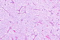
ChRCC - low mag. (WC/Nephron)
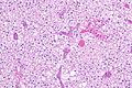
ChRCC - intermed. mag. (WC/Nephron)
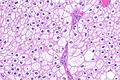
ChRCC - high mag. (WC/Nephron)
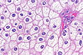
ChRCC - very high mag. (WC/Nephron)
ChRCC - high mag. (WC/Nephron)
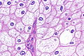
ChRCC - very high mag. (WC/Nephron)


Eosinophilic variant
Features - eosinophilic variant:[4]
- Eosinophilic (finely granular) cytoplasm.
- Perinuclear clearing - key feature.
- Periphery of cell distinct.
- Smaller cells than classic subtype.
Notes:
- May have psammoma bodies.
DDx:
- Oncocytoma - particularly the eosinophilic variant.
- IHC may be useful to differentiate (CK7: oncocytoma = cytoplasm +ve, chromophobe = cell membrane +ve).
- A comparison based on histomorphology: Tabular comparison between ChRCC & oncocytoma.
- Oncocytoma typically has: no perinuclear clearing, no raisinoid nuclei, no binucleation.
- Clear cell RCC, eosinophilic variant.
- Perinuclear clearing is not seen in clear cell RCC.
- ChRCC has wisps in the cytoplasm.
- Renal hybrid oncocytic/chromophobe tumour.
- Other renal tumours with eosinophilic cytoplasm.
Images
Case 1
Eosinophilic chromophobe RCC. (WC/Nephron)

Case 2
EVChRCC - low mag.
EVChRCC - intermed. mag.
EVChRCC - intermed. mag.
EVChRCC - high mag.
EVChRCC - very high mag.
EVChRCC - very high mag.


www
Stains
- Hale's colloidal iron +ve (blue granular cytoplasmic).
- Oncocytoma -ve.
Note:
- This seems to be a difficult stain to get working.
Images
- ChRCC Hale's colloidal iron - several images (nature.com).
- ChRCC Hale's colloidal iron (ultrapath.org).[8]
- ChRCC Hale's colloidal iron (diagnosticpathology.org).
IHC
- CK7 +ve cell membrane.[4]
- Useful for differentiating from oncocytoma.
- CD117 +ve.
- Vimentin -ve.[9][10]
Others:[11]
- CD10 -ve.
- RCC -ve.
Uncommon stains for ChRCC versus oncocytoma:
ChRCC versus clear cell RCC
- CD117 +ve.
- -ve in CCRCC.
- CA9 (carbonic anhydrase 9) -ve.
- +ve (strong membranous) in CCRCC.[15]
- CK7 +ve.
- -ve in CCRCC.
Others:
- DOG1 +ve (Swalchick et al.[16] 32 +ve of 37 cases; Zhao et al.[17] 32 +ve of 32 cases).
- Useful in ChRCC versus clear cell RCC.
A panel:
- CK7, PAX8, CD117, CD10, Mueller-Mowry stain.
Comparison between some renal tumours with eosinophilic cytoplasm
| Tumour | CK7 | CD117 | GATA3 |
|---|---|---|---|
| Renal oncocytoma | -ve † | +ve | -ve/+ve |
| Chromophobe renal cell carcinoma | +ve ‡ | +ve | -ve |
| Low-grade oncocytic tumour | +ve | -ve | +ve |
† may have scattered positive cells
‡ diffuse and strong
Molecular
- Extensive aneusomy (monosomy?):[18]
- Loss of chromosomes: 1, 2, 6, 10, 13, 17, 21.
EM
Ultrastructural features:[19]
- Microvesicles.
- Complex plicated cell membrane.
Images
Sign out
KIDNEY, RIGHT UPPER POLE, PARTIAL NEPHRECTOMY:
- CHROMOPHOBE RENAL CELL CARCINOMA.

COMMENT:
The sections show a mix of clear cells with wispy cytoplasm, and cells with eosinophilic
cytoplasm and perinuclear halos. There are no true papillae.

Stains and immunostains:
Positive: CK7, CAM5.2, EMA, pankeratin, CD117, colloidal iron.
Negative: AMACR, CD10, CD68, RCC, vimentin.
Micro
The sections show a sheeting tumour with abundant and eosinophilic cytoplasm. The tumour cell cell membranes are prominent and plant-like. Perinuclear clearing is present. Binucleation is frequent and the nuclear membranes moderately irregular. No definite papillae or macrophages are present. Nucleoli are not prominent. Small blood vessels are not prominent.
Alternate
The sections show a tumour composed of compact nests. The cytoplasm is abundant and eosinophilic. Perinuclear clearing is prominent. Binucleation is frequent and the nuclear membranes moderately irregular to round. No definite papillae or macrophages are present. Nucleoli are not prominent.
See also
References
- ↑ Delahunt, B.; Sika-Paotonu, D.; Bethwaite, PB.; McCredie, MR.; Martignoni, G.; Eble, JN.; Jordan, TW. (Jun 2007). "Fuhrman grading is not appropriate for chromophobe renal cell carcinoma.". Am J Surg Pathol 31 (6): 957-60. doi:10.1097/01.pas.0000249446.28713.53. PMID 17527087.
- ↑ Humphrey, Peter A; Dehner, Louis P; Pfeifer, John D (2008). The Washington Manual of Surgical Pathology (1st ed.). Lippincott Williams & Wilkins. pp. 290. ISBN 978-0781765275.
- ↑ Kuroda, N.; Tanaka, A.; Ohe, C.; Mikami, S.; Nagashima, Y.; Sasaki, T.; Inoue, K.; Hes, O. et al. (Nov 2012). "Review of renal oncocytosis (multiple oncocytic lesions) with focus on clinical and pathobiological aspects.". Histol Histopathol 27 (11): 1407-12. PMID 23018240.
- ↑ 4.0 4.1 4.2 4.3 Zhou, Ming; Magi-Galluzzi, Cristina (2006). Genitourinary Pathology: A Volume in Foundations in Diagnostic Pathology Series (1st ed.). Churchill Livingstone. pp. 293. ISBN 978-0443066771.
- ↑ Kuroda, N.; Toi, M.; Hiroi, M.; Enzan, H. (Jan 2003). "Review of chromophobe renal cell carcinoma with focus on clinical and pathobiological aspects.". Histol Histopathol 18 (1): 165-71. PMID 12507296.
- ↑ Siracusano, S.; Novara, G.; Antonelli, A.; Artibani, W.; Bertini, R.; Carini, M.; Carmignani, G.; Ciciliato, S. et al. (Dec 2012). "Prognostic role of tumour multifocality in renal cell carcinoma.". BJU Int 110 (11 Pt B): E443-8. doi:10.1111/j.1464-410X.2012.11121.x. PMID 22502873.
- ↑ Cotran, Ramzi S.; Kumar, Vinay; Fausto, Nelson; Nelso Fausto; Robbins, Stanley L.; Abbas, Abul K. (2005). Robbins and Cotran pathologic basis of disease (7th ed.). St. Louis, Mo: Elsevier Saunders. pp. 1016-7. ISBN 0-7216-0187-1.
- ↑ URL: http://www.ultrapath.org/oldsite/cases99/sep99/cotm9-2.html. Accessed on: 9 October 2011.
- ↑ Zhang, W.; Yu, WJ.; Jiang, YX.; Li, YJ.; Han, F.; Liu, Y.; Han, ZL. (Feb 2012). "[Chromophobe renal cell carcinoma: a clinicopathologic study and immunophenotypes of 42 cases].". Zhonghua Bing Li Xue Za Zhi 41 (2): 76-80. PMID 22455881.
- ↑ Din, NU.; Fatima, S.; Ahmad, Z. (Dec 2013). "Chromophobe renal cell carcinoma: a morphologic and immunohistochemical study of 45 cases.". Ann Diagn Pathol 17 (6): 508-13. doi:10.1016/j.anndiagpath.2013.06.005. PMID 24095630.
- ↑ Geramizadeh, B.; Ravanshad, M.; Rahsaz, M.. "Useful markers for differential diagnosis of oncocytoma, chromophobe renal cell carcinoma and conventional renal cell carcinoma.". Indian J Pathol Microbiol 51 (2): 167-71. PMID 18603673.
- ↑ Memeo L, Jhang J, Assaad AM, et al. (February 2007). "Immunohistochemical analysis for cytokeratin 7, KIT, and PAX2: value in the differential diagnosis of chromophobe cell carcinoma". Am. J. Clin. Pathol. 127 (2): 225–9. doi:10.1309/9KWEA4W9Y94D1AEE. PMID 17210525. http://ajcp.ascpjournals.org/cgi/pmidlookup?view=long&pmid=17210525.
- ↑ Jain, S.; Roy, S.; Amin, M.; Acquafondata, M.; Yin, M.; Laframboise, W.; Bastacky, S.; Pantanowitz, L. et al. (Dec 2013). "Amylase α-1A (AMY1A): A Novel Immunohistochemical Marker to Differentiate Chromophobe Renal Cell Carcinoma From Benign Oncocytoma.". Am J Surg Pathol 37 (12): 1824-30. doi:10.1097/PAS.0000000000000108. PMID 24225843.
- ↑ Amin MB, Epstein JI, Ulbright TM, et al. (August 2014). "Best practices recommendations in the application of immunohistochemistry in urologic pathology: report from the international society of urological pathology consensus conference". Am. J. Surg. Pathol. 38 (8): 1017–22. doi:10.1097/PAS.0000000000000254. PMID 25025364.
- ↑ Al-Ahmadie HA, Alden D, Fine SW, et al. (July 2011). "Role of immunohistochemistry in the evaluation of needle core biopsies in adult renal cortical tumors: an ex vivo study". Am. J. Surg. Pathol. 35 (7): 949–61. doi:10.1097/PAS.0b013e31821e25cd. PMID 21677535.
- ↑ Swalchick, W.; Shamekh, R.; Bui, MM. (Oct 2015). "Is DOG1 Immunoreactivity Specific to Gastrointestinal Stromal Tumor?". Cancer Control 22 (4): 498-504. PMID 26678977.
- ↑ Zhao, W.; Tian, B.; Wu, C.; Peng, Y.; Wang, H.; Gu, WL.; Gao, FH. (Apr 2015). "DOG1, cyclin D1, CK7, CD117 and vimentin are useful immunohistochemical markers in distinguishing chromophobe renal cell carcinoma from clear cell renal cell carcinoma and renal oncocytoma.". Pathol Res Pract 211 (4): 303-7. doi:10.1016/j.prp.2014.12.014. PMID 25596994.
- ↑ Humphrey, Peter A; Dehner, Louis P; Pfeifer, John D (2008). The Washington Manual of Surgical Pathology (1st ed.). Lippincott Williams & Wilkins. pp. 292. ISBN 978-0781765275.
- ↑ 19.0 19.1 Lee, W. (Apr 2011). "Imprint cytology of the chromophobe renal cell carcinoma: Correlation with the histological and ultrastructural features.". J Cytol 28 (2): 77-80. doi:10.4103/0970-9371.80749. PMID 21713152.