Difference between revisions of "Chondromyxoid fibroma"
Jump to navigation
Jump to search
| Line 1: | Line 1: | ||
{{ Infobox diagnosis | |||
| Name = {{PAGENAME}} | |||
| Image = | |||
| Width = | |||
| Caption = | |||
| Synonyms = | |||
| Micro = spindle cells or stellate cells in a myxoid or chondroid stroma, lobules with hypocellular centers and hypercellular peripheries, +/-[[giant cells]] in the hypercellular periphery, scattered calcifications, no true hyaline cartilage formation, no mitotic activity | |||
| Subtypes = | |||
| LMDDx = [[chondroblastoma]], [[chondrosarcoma]], [[phosphaturic mesenchymal tumour]] (case report) | |||
| Stains = | |||
| IHC = | |||
| EM = | |||
| Molecular = | |||
| IF = | |||
| Gross = | |||
| Grossing = | |||
| Site = [[bone]] ([[metaphysis]]) - see ''[[bone tumours]]'' | |||
| Assdx = | |||
| Syndromes = | |||
| Clinicalhx = teenager/young adult | |||
| Signs = | |||
| Symptoms = | |||
| Prevalence = uncommon | |||
| Bloodwork = | |||
| Rads = | |||
| Endoscopy = | |||
| Prognosis = benign | |||
| Other = | |||
| ClinDDx = | |||
| Tx = | |||
}} | |||
'''Chondromyxoid fibroma''' is a rare benign [[Chondro-osseous tumours|chondro-osseous tumour]] typically found in the [[metaphysis]] of teenagers or young adults. | '''Chondromyxoid fibroma''' is a rare benign [[Chondro-osseous tumours|chondro-osseous tumour]] typically found in the [[metaphysis]] of teenagers or young adults. | ||
| Line 20: | Line 51: | ||
DDx: | DDx: | ||
*[[Chondroblastoma]]. | *[[Chondroblastoma]]. | ||
**Likewise has immature cartilage but (1) epiphyseal location, (2) chickenwire-like calcifications. | **Likewise has immature cartilage but (1) [[epiphysis|epiphyseal]] location, (2) chickenwire-like calcifications. | ||
*[[Chondrosarcoma]]. | *[[Chondrosarcoma]]. | ||
**Different age group. | **Different age group. | ||
**Mature hyaline cartilage formation. | **Mature hyaline [[cartilage]] formation. | ||
**Tumour permeation of the surrounding bone. | **Tumour permeation of the surrounding bone. | ||
**Mitotic activity. | **Mitotic activity. | ||
*[[Phosphaturic mesenchymal tumour]] - case report.<ref>{{Cite journal | last1 = Suryawanshi | first1 = P. | last2 = Agarwal | first2 = M. | last3 = Dhake | first3 = R. | last4 = Desai | first4 = S. | last5 = Rekhi | first5 = B. | last6 = Reddy | first6 = KB. | last7 = Jambhekar | first7 = NA. | title = Phosphaturic mesenchymal tumor with chondromyxoid fibroma-like feature: an unusual morphological appearance. | journal = Skeletal Radiol | volume = 40 | issue = 11 | pages = 1481-5 | month = Nov | year = 2011 | doi = 10.1007/s00256-011-1159-6 | PMID = 21533894 }}</ref> | |||
===Images=== | ===Images=== | ||
Revision as of 03:57, 14 November 2014
| Chondromyxoid fibroma | |
|---|---|
| Diagnosis in short | |
|
| |
| LM | spindle cells or stellate cells in a myxoid or chondroid stroma, lobules with hypocellular centers and hypercellular peripheries, +/-giant cells in the hypercellular periphery, scattered calcifications, no true hyaline cartilage formation, no mitotic activity |
| LM DDx | chondroblastoma, chondrosarcoma, phosphaturic mesenchymal tumour (case report) |
| Site | bone (metaphysis) - see bone tumours |
|
| |
| Clinical history | teenager/young adult |
| Prevalence | uncommon |
| Prognosis | benign |
Chondromyxoid fibroma is a rare benign chondro-osseous tumour typically found in the metaphysis of teenagers or young adults.
General
- Uncommon and benign.[1]
- Teenagers or young adults.
Gross
- Metaphyseal lesion - classic location.[2]
- Well-circumscribed.
- Fragments of white-grey rubbery tissue.
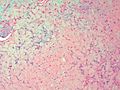
Microscopic
Features:[3]
- Spindle cells or stellate cells in a myxoid or chondroid stroma.
- Lobules with hypocellular centers and hypercellular peripheries.
- Giant cells in the hypercellular periphery.
- Scattered calcifications.
- No true hyaline cartilage formation.
- No mitotic activity.
DDx:
- Chondroblastoma.
- Likewise has immature cartilage but (1) epiphyseal location, (2) chickenwire-like calcifications.
- Chondrosarcoma.
- Different age group.
- Mature hyaline cartilage formation.
- Tumour permeation of the surrounding bone.
- Mitotic activity.
- Phosphaturic mesenchymal tumour - case report.[4]
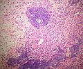
Images
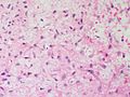
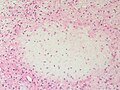
Stellate cells in a myxoid stroma. (SKB)
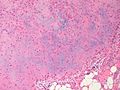
Lobule with calcification. (SKB)
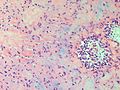
Stellate cells in a myxohyaline stroma. (SKB)
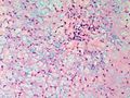
Stellate cells in a myxoid stroma. (SKB)
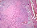
Lobule with a darker (hypercellular) periphery and lighter (hypocellular) center. (SKB)
Stellate cells in a myxohyaline stroma. (SKB)
Stellate cells in a myxohyaline stroma. (SKB)
Stellate cells in a myxoid stroma. (SKB)
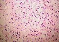
Stellate cells in a myxoid stroma. (SKB)

Low power view displaying the lobular nature of these proliferations. (SKB)
Focus of amorphous calcification. (SKB)
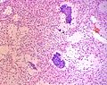
Foci of amorphous calcification. (SKB)
.jpg)


www:
- Chondromyxoid fibroma - low mag. (webpathology.com).
- Chondromyxoid fibroma - high mag. (webpathology.com).
- Tumor Library [1].
- Tumor Library [2].
- Tumor Library [3].
- Tumor Library [4].
- Tumor Library [5].
- Tumor Library [6].
- Diagnostic Pathology [7].
![[1]](http://www.tumorlibrary.com/case/images/1490.jpg)
![[2]](http://www.tumorlibrary.com/case/images/1491.jpg)
![[3]](http://www.tumorlibrary.com/case/images/3618.jpg)
![[4]](http://www.tumorlibrary.com/case/images/3619.jpg)
![[5]](http://www.tumorlibrary.com/case/images/3621.jpg)
![[6]](http://www.tumorlibrary.com/case/images/3623.jpg)
![[7]](http://www.diagnosticpathology.org/content/figures/1746-1596-2-44-4.jpg)
Molecular
- Activating rearrangements of GRM1 (metabotropic glutamate receptor 1).[5]
See also
References
- ↑ Bhamra, JS.; Al-Khateeb, H.; Dhinsa, BS.; Gikas, PD.; Tirabosco, R.; Pollock, RC.; Skinner, JA.; Aston, WJ. et al. (2014). "Chondromyxoid fibroma management: a single institution experience of 22 cases.". World J Surg Oncol 12: 283. doi:10.1186/1477-7819-12-283. PMID 25217119.
- ↑ Budny, AM.; Ismail, A.; Osher, L.. "Chondromyxoid fibroma.". J Foot Ankle Surg 47 (2): 153-9. doi:10.1053/j.jfas.2007.08.013. PMID 18312923.
- ↑ Humphrey, Peter A; Dehner, Louis P; Pfeifer, John D (2008). The Washington Manual of Surgical Pathology (1st ed.). Lippincott Williams & Wilkins. pp. 642. ISBN 978-0781765275.
- ↑ Suryawanshi, P.; Agarwal, M.; Dhake, R.; Desai, S.; Rekhi, B.; Reddy, KB.; Jambhekar, NA. (Nov 2011). "Phosphaturic mesenchymal tumor with chondromyxoid fibroma-like feature: an unusual morphological appearance.". Skeletal Radiol 40 (11): 1481-5. doi:10.1007/s00256-011-1159-6. PMID 21533894.
- ↑ Nord, KH.; Lilljebjörn, H.; Vezzi, F.; Nilsson, J.; Magnusson, L.; Tayebwa, J.; de Jong, D.; Bovée, JV. et al. (May 2014). "GRM1 is upregulated through gene fusion and promoter swapping in chondromyxoid fibroma.". Nat Genet 46 (5): 474-7. doi:10.1038/ng.2927. PMID 24658000.