Difference between revisions of "Chondro-osseous tumours"
(re-arr) |
m (→Brown tumour: format) |
||
| Line 604: | Line 604: | ||
*Drugs (thiazides ... others). | *Drugs (thiazides ... others). | ||
=== | ===Microscopic=== | ||
Features: | Features: | ||
*Fibrosis. | *Fibrosis. | ||
Revision as of 01:41, 28 July 2011
Chondro-osseous tumours occasionally cross the desk of the pathologist. They are grouped together as bone may develop from cartilage.
Primary bone tumours are rare; the most common bone tumour is metastases.[1]
Bone tumours occasionally are lumped with soft tissue tumours. Soft tissue tumours are dealt with in the soft tissue lesions article. An introduction to bone is found in the bone article. An introduction to cartilage is found in the cartilage article.
General
- Diagnosis of a primary bone tumour should not be made without radiologic & clinical information!
- Metastasis:primary bone tumours = >20:1.[1]
Common malignant
- Osteosarcoma.
- Chondrosarcoma.
- Ewing's sarcoma.
- Multiple myeloma.
- Metastases.
- Most common tumours metastatic to bone (mnemonic: BLT with Ketchup & Pickles):
Epidemiology:[2]
- Osteosarcoma -> 2nd decade.
- Ewing's ->5-20 yrs.
- Chondrosarcoma -> from enchondroma or osteochrondroma -- patients over 40 yrs.
- Multiple myeloma -> most common primary bone tumour in adults.
Malignant bone tumours by age
Most common by age:[3]
- <1 year old - neuroblastoma.
- 1-10 years old - Ewing's of tubular bones.
- 10-30 years old - osteosarcoma, Ewing's of flat bones.
- 30-40 years old - reticulum cell sarcoma, fibrosarcoma, parosteal osteosarcoma, malignant giant cell tumour, lymphoma.
- >40 years old - mets, multiple myeloma, chondrosarcoma.
Benign aggressive bone tumours
- Giant cell tumours.
- Osteoblastoma.
- Thought to be related to osteoid osteoma.
- If in long bones often diaphyseal.
Summary tables
Bone tumours
| Entity | Key feature | Other features | Radiology / gross | Clinical | Stains / other | Image |
| Osteoid osteoma | osteoblastic rimming | anastomosing bony trabeculae | <= 1.5 cm | painful, NSAIDs remove pain, young | IHC / other | low mag., high mag. |
| Osteoblastoma | osteoblastic rimming | anastomosing bony trabeculae | > 1.5 cm | not painful | IHC / other | low mag., high mag. |
| Ewing sarcoma | small round blue cell tumour | cytoplasmic clearing (due to glycogen) | Radiology / gross | pediatric | PAS+, PASD- | intermed. mag., high mag. - PAS |
| Osteosarcoma | osteoid | Other features | Radiology / gross | Clinical | no stains; many subtypes | very high mag. |
| Giant cell tumour of bone | abundant giant cells | nuclei of surrounding cells similar to those in giant cells | Radiology / gross | Clinical | IHC / other | high mag. |
Cartilage tumours
| Entity | Key feature | Other features | Radiology / gross | Clinical | Stains / other | Image |
| Enchondroma | Key feature | Other features | Radiology / gross | Clinical | IHC / other | Image |
| Chondroblastoma | Key feature | Other features | Radiology / gross | Clinical | IHC / other | Image |
| Chondrosarcoma | Key feature | Other features | Radiology / gross | Clinical | IHC / other | Image |
Other
| Entity | Key feature | Other features | Radiology / gross | Clinical | Stains / other | Image |
| Osteochondroma | Key feature | Other features | Radiology / gross | Clinical | IHC / other | Image |
| Adamantinoma | bisphasic - stroma & epithelium | Other features | Radiology / gross | Clinical | IHC / other | Image |
| Diffuse tenosynovial giant-cell tumour (AKA PVNS) | pigmented giant cells | nodules | Radiology / gross | Clinical | IHC / other | Image |
| Brown tumour | Key feature | Other features | Radiology / gross | due to hypercalcemia; not a neoplasm | IHC / other | Image |
Cartilage
Enchondroma
General
- Benign thingy.
- Usu. legs and feet.
- May be difficult to separate from chondrosarcoma.
- Multiple chondromas = enchondromatosis; three distinct syndromes.[6]
Radiology:[6]
- Lytic lesion.
- Usu. close to a growth plate.
Clinical:[6]
- Pain.
Microscopic
Features:
- Ctyologically benign cells is spaced nests.
Images:
Chondroblastoma
General
- Growth plate lesion.
- Sclerotic margin.
- "Young" = growth plates open.
Microscopic
Features:[7]
- Abundant extracellular material - pink on H&E stain - looks vaguely like cartilage.
- Chondroblasts:
- Nuclear morphology variable: ovoid, folded or grooved.
- Moderate-abundant eosinophilic cytoplasm.
- +/-Calcifications surround cells nests ("chickenwire" appearance) - classic feature.
- +/-Giant cells.
- May lead to confusion with giant cell tumour.
Images:
- Chondroblastoma - intermed. mag. (WC).
- Chondroblastoma - very high mag. (WC).
- Chondroblastoma (medscape.com).[8]
- Chondroblastoma with "chickenwire" appearance (medscape.com).[8]
IHC
Features:[7]
- S100 +ve.
- Vimentin +ve.[8]
Chondrosarcoma
General
- Usually a good prognosis.
Clinical/epidemiologic features:[9]
- Usually arise in a (benign) abnormality of cartilage (e.g. osteochondroma, enchondroma).
- May be associated with a syndrome:
- Olier disease (multiple enchondromatosis).
- Maffucci syndrome (multiple enchondromas and hemangiomas).
Notes:
- Review article (from oncology perspective): PMID 17545802.
Subtypes
Several subtypes exist:
- Chondrosarcoma not otherwise specified (NOS).
- Juxtacortical chondrosarcoma.
- Myxoid chondrosarcoma.
- Mesenchymal chondrosarcoma.
- Clear cell chondrosarcoma.
- Dedifferentiated chondrosarcoma chondrosarcoma.
Microscopic
- "Abnormal cartilage":
- +/-Nuclear atypia - high grade lesions.
- High grade lesions:
- Nuclear clearing.
- Nucleoli.
- Hyperchromasia.
- Low/intermediate grade lesions:
- Bi-nucleation.
- Hypochromatic enlarged nuclei.
- Infiltration of lamellar bone ("invasion") - not common - diagnostic.
- High grade lesions:
- Increased cellularity.
- More cellular than cartilage... but relatively paucicellular compared to other sarcomas.
- Irregular spacing of chondrocytes.
- +/-Nuclear atypia - high grade lesions.
Notes:
- Low grade chondrosarcoma are not cytologically malignant; the diagnosis rests mostly on radiologic findings.
- The exception is infiltration of lamellar bone -- this is diagnostic of chondrosarcoma.[12]
Images:
.jpg)
.jpg)
.jpg)
DDx:
Variants
Mesenchymal chondrosarcoma
- Arise in soft tissue; this is where the name comes from.[13]
- Rare variant of chondrosarcoma.
Microscopic: Features:
- "White clouds in a blue sky".
Image:
Myxoid chondrosarcoma
Microscopic: Features:
- Chordoma-like:
- Myxoid background.
- Small cells with eosinophilic cytoplasm.
DDx:
Image:
Grading
Features:[16]
- Grade I: mild-to-moderate increase of cellularity +/- binucleated cells.
- Grade II: between Grade I and Grade III.
- Grade III: nuclear pleomorphism, mitoses common.
IHC
- S-100 -ve. (???)
Bone
Osteoid osteoma
General
- Benign bone lesion.
Clinical:[17]
- Extremely painful.
- Relieved by NSAIDS.
Microscopic
Features:[17]
- Anastomosing bony trabeculae with:
- Variable mineralization.
- Mineralization (calcium phosphate) = purple on H&E stain.
- Osteoblasts rimming.
- Cells line-up at edge of bone.
- Variable mineralization.
Images:
- Osteoid osteoma - CT scan (med.utah.edu).
- Osteoid osteoma (sciencephoto.com).
- Osteoid osteoma - low mag. (WC).
- Osteoid osteoma - high mag. (WC).
Notes:
- Histomorphologically near identical/indistinguishable from osteoblastoma.[18]
Osteoblastoma
General
- Benign bone tumour.
Microscopic
Features:[17]
- Anastomosing bony trabeculae with:
- Osteoblasts rimming.
- Cells line-up at edge of bone.
- Osteoblasts rimming.
Notes:
- Histomorphologically near identical/indistinguishable from osteoid osteoma.[18]
- Must be greater 1.5 cm by definition.[18]
Images:
Ewing sarcoma
- AKA EWS/pPNET, AKA (confusingly) EWS/PNET:
- EWS = Ewing sarcoma.
- pPNET = peripheral primitive neuroectodermal tumour.
- EWS and pPNET were once thought to be different tumours.
Notes:
- Peripheral primitive neuroectodermal tumour should not be confused with primitive neuroectodermal tumour, commonly abbreviated PNET, a (supertentorial) brain tumour with similarities to medulloblastoma.
General
Clinical:
- Painful.
- Usually younger than 20 years.
- Second most common malignant bone tumour in children.
- Most common malignant bone tumour = osteosarcoma (AKA osteogenic sarcoma).
Poor prognostic factors:[19]
- Age (18 years-old+).
- Pelvis (extremity = good).
- >8 cm.
- Metastases.
- EWS-FL1 fusion type 2.
- >90% necrosis.
Etiology:
- Unknown origin; hypothesis: Ewing sarcoma arises from mesenchymal stem cell.[20]
Radiology
Features:[21]
- Long bones, diaphyses.
- Destructive.
- "Onion-skin" periosteal reaction.
Microscopic
Features:[22]
- Scant clear cytoplasm (contain glycogen -- PAS +ve, PAS-D -ve) - key feature.
- Round small nucleus.
- Usu. lack nucleoli.
- Usu. minimal-moderate size variation.
- Mitoses (common).
Notes:
- It is a small round cell tumour.
- Rhabdomyosarcoma occasionally has cytoplasmic clearing (due to glycogen).[23]
Images:
IHC
Features:[24]
- CD99 +ve -- 1. diffuse, 2. plasma membrane staining; both required -- most specific.
- FLI-1 +ve.[25]
- CD45 -ve.
- Done to r/o lymphoma.
- +/-Neural markers (NSE, synaptophysin, CD57 (??? CD56 ???), S100).
- +/-Cytokeratins.
- Caveolin-1 +ve in ~ 85% of EWS.[26]
Notes:[27]
- CD99 +ve
- Plasma membrane staining tumours:
- Lymphoblastic lymphoma/leukemia.
- Angiomatoid fibrous histiocytoma.
- Desmoplastic small round cell tumour.
- Weak/cytoplasmic staining:
- Plasma membrane staining tumours:
- FLI-1 +ve:[25]
- Vascular neoplasms.
- -/+Merkel cell carcinoma.
- -/+Melanoma.
Molecular diagnostics
Common features:
- EWS/FLI-1 fusion gene formation due to translocation: t(11;22)(q24;q12).[28][29]
- Often detected by RT-PCR (with EWS 5' and FLI-1 3' primers).
- Type 1 = EWS exon 7 + FLI-1 exon 6; good prognosis.
- Type 2 = others; poor prognosis.
Notes:
- The t(11;22)(q24;q12) is seen in ~90% of EWS/PNET... but also in:
- Olfactory neuroblastoma.
- Small cell osteogenic sarcoma (small cell variant of osteosarcoma).
- Polyphenotypic tumours.
- Rhabdomyosarcoma.
- Neuroblastoma (possibly).
- Several other translocations exist.
- Lack of molecular findings does not exclude Ewing sarcoma.
- Testing:
- A break apart probe for EWS is a common way to look for pathologic change, as it covers almost all variants.
Electron microscopy
- Primitive cell junctions.
- Clear zone (glycogen lakes).
Osteosarcoma
- AKA osteogenic sarcoma.
General
- Most common malignant bone tumour in children.
Trivia:
- Terry Fox was afflicited by this tumour.
Definition
- Tumour that makes osteoid.
- Osteoid = (extracellular) organic component of bone, normally produced by osteoblasts (cells which make bone matrix).
Microscopic
Features:
- Cells with malignant features (e.g. nuclear membrane irregularities, marked nuclear size differences, mitoses) surrounded by delicate strands of osteoid.
- Osteoid on H&E: pink, homogenous, "glassy".
- Tumours typically very cellular - when compared to normal bone.
- Large (multinucleated) osteoclast-like giant cells may be seen.[30]
Images:
Subtypes
- Many subtypes exist.
- Conventional osteosarcoma (high grade).
- Osteoblastic.
- Fibroblastic - undifferentiated pleomorphic sarcoma-like/MFH-like.
- Chondroblastic - may be confused with chondrosarcoma.
- Small cell - may mimic (other) small round cell tumours.
- Telangiectatic - extremely vascular.
- Parosteal.
- Low grade.
- Arises from surface of bone.[33]
- Periosteal.
- Low grade central.
- High grade surface.
- Secondary - arise due to something else (e.g. Paget disease of the bone (~80% of secondary osteosarcomas), radiation (~15% of secondary osteosarcomas)).[34]
- Gnathic - jaw bones; usu. chondroblastic.
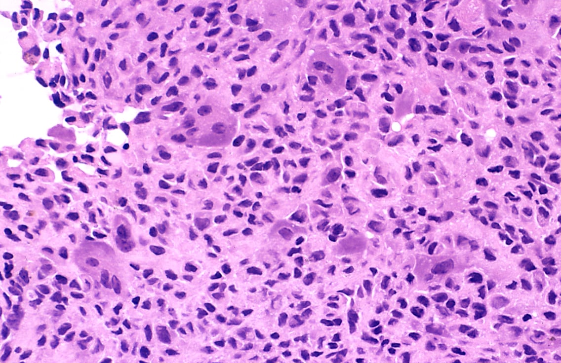
Giant cell tumour of bone
General
Features:[35]
- Approximately 5% of primary bone tumours.
- Typical age: 20-45 years.
Clinical
- May present with joint pain, immobility.
Note:
- Several types of giant cell tumours exist.
Microscopic
Features:[36]
- Giant cells with abundant nuclei (usu. >10 in the plane of section).
- Usu. have prominent nucleoli.
- Mononuclear cells and small multinucleated cells with nuclei similar to those in the giant cells - key feature.
Images:
IHC
Other
This section collects stuff that doesn't neatly fit into the bone or cartilage category.
Osteochondroma
General
- Benign metaphyseal lesions.
- Very common.
- Abnormal outgrowth of bone and cartilage.
Microscopic
Features:
- Normal bone and cartilage.
Diffuse tenosynovial giant-cell tumour
- AKA tenosynovial giant-cell tumour, diffuse type.
- Previously known as pigmented villonodular synovitis (PVNS).[39]
General
- Course: benign.
- Giant cell tumor of the tendon sheath is considered to be the soft-tissue counterpart of PVNS.[40]
Microscopic
Features:[41]
- Subsynovial nodules composed of cells with:
- Abundant cytoplasm.
- Pale nuclei.
- Multinucleated giant cells.
- Hemosiderin-laden macrophages.
- Foam cells.
Images:
Adamantinoma
General
Features:[21]
- Rare: < 1% of bone tumours.
- 25-35 years old.
- Tibia, fibula.
- Benign, may be locally aggressive.
- Cousin of ameloblastoma. (???)
Radiology
- Intracortical, radiolucent.
Microscopic
Features:
- Biphasic tumour:
- Fibrous/spindle cell component.
- Epithelial component.
Images:
DDx:[43]
- Vascular tumours (Epithelioid hemangioendothelioma).
- Metastatic carcinoma.
IHC
Features:[43]
- CK14 +ve (HMWK).[44]
- CK19 +ve (LMWK).
- CK8/18 -ve (LMWK).
Brown tumour
General
- Not a true neoplasm,[45] i.e. the name is a misnomer.
- May (clinically) mimic a true neoplasm.
- Due to hyperparathyroidism - usually parathyroid adenoma.
- Usually secondary to chronic renal failure.
Hypercalcemia DDx
Mnemonic GRIMED:[46]
- Granulomatous disease (tuberculosis, sarcoidosis).
- Renal disease.
- Immobility.
- Malignancy (esp. squamous cell carcinoma, plasmacytoma).
- Endocrine (primary hyperparathyroidism - leads to brown cell tumour).
- Drugs (thiazides ... others).
Microscopic
Features:
- Fibrosis.
- +/-Giant cells.
DDx:
- Giant cell tumour of bone and other giant cell lesions.
See also
References
- ↑ 1.0 1.1 Humphrey, Peter A; Dehner, Louis P; Pfeifer, John D (2008). The Washington Manual of Surgical Pathology (1st ed.). Lippincott Williams & Wilkins. pp. 632. ISBN 978-0781765275.
- ↑ TN05 OR42.
- ↑ TN05 OR42.
- ↑ TN05 OR41.
- ↑ URL: http://www.emedicine.com/RADIO/topic494.htm.
- ↑ 6.0 6.1 6.2 URL: http://emedicine.medscape.com/article/389224-overview. Accessed on: 25 December 2010.
- ↑ 7.0 7.1 Humphrey, Peter A; Dehner, Louis P; Pfeifer, John D (2008). The Washington Manual of Surgical Pathology (1st ed.). Lippincott Williams & Wilkins. pp. 642. ISBN 978-0781765275.
- ↑ 8.0 8.1 8.2 URL: http://emedicine.medscape.com/article/1254949-diagnosis. Accessed on: 31 December 2010.
- ↑ Skubitz KM, D'Adamo DR (November 2007). "Sarcoma". Mayo Clin. Proc. 82 (11): 1409–32. PMID 17976362. http://www.mayoclinicproceedings.com/content/82/11/1409.long.
- ↑ IAV. 26 February 2009.
- ↑ Klatt, Edward C. (2006). Robbins and Cotran Atlas of Pathology (1st ed.). Saunders. pp. 417. ISBN 978-1416002741.
- ↑ BD. 28 April 2011.
- ↑ Dowling EA (June 1964). "Mesenchymal chondrosarcoma". J Bone Joint Surg Am 46: 747–54. PMID 14161087. http://www.ejbjs.org/cgi/reprint/46/4/747.pdf.
- ↑ Fisher C (May 2000). "Parachordoma exists--but what is it?". Adv Anat Pathol 7 (3): 141–8. PMID 10809219.
- ↑ URL: http://www.cttr.org/cms/?p=736. Accessed on: 1 May 2011.
- ↑ Humphrey, Peter A; Dehner, Louis P; Pfeifer, John D (2008). The Washington Manual of Surgical Pathology (1st ed.). Lippincott Williams & Wilkins. pp. 643. ISBN 978-0781765275.
- ↑ 17.0 17.1 17.2 Mills, Stacey E; Carter, Darryl; Greenson, Joel K; Oberman, Harold A; Reuter, Victor E (2004). Sternberg's Diagnostic Surgical Pathology (4th ed.). Lippincott Williams & Wilkins. pp. 285. ISBN 978-0781740517.
- ↑ 18.0 18.1 18.2 Mills, Stacey E; Carter, Darryl; Greenson, Joel K; Oberman, Harold A; Reuter, Victor E (2004). Sternberg's Diagnostic Surgical Pathology (4th ed.). Lippincott Williams & Wilkins. pp. 286. ISBN 978-0781740517.
- ↑ PST. 14 February 2011.
- ↑ Lin PP, Wang Y, Lozano G (2011). "Mesenchymal Stem Cells and the Origin of Ewing's Sarcoma". Sarcoma 2011. doi:10.1155/2011/276463. PMC 2952797. PMID 20953407. https://www.ncbi.nlm.nih.gov/pmc/articles/PMC2952797/.
- ↑ 21.0 21.1 Humphrey, Peter A; Dehner, Louis P; Pfeifer, John D (2008). The Washington Manual of Surgical Pathology (1st ed.). Lippincott Williams & Wilkins. pp. 650. ISBN 978-0781765275.
- ↑ PST. 22 February 2010.
- ↑ PST. 14 February 2011.
- ↑ Humphrey, Peter A; Dehner, Louis P; Pfeifer, John D (2008). The Washington Manual of Surgical Pathology (1st ed.). Lippincott Williams & Wilkins. pp. 651. ISBN 978-0781765275.
- ↑ 25.0 25.1 Rossi S, Orvieto E, Furlanetto A, Laurino L, Ninfo V, Dei Tos AP (May 2004). "Utility of the immunohistochemical detection of FLI-1 expression in round cell and vascular neoplasm using a monoclonal antibody". Mod. Pathol. 17 (5): 547–52. doi:10.1038/modpathol.3800065. PMID 15001993. http://www.nature.com/modpathol/journal/v17/n5/full/3800065a.html.
- ↑ PST. 14 February 2011.
- ↑ PST. 22 February 2010.
- ↑ URL: http://atlasgeneticsoncology.org/Tumors/Ewing5010.html. Accessed on: 23 February 2010.
- ↑ Turc-Carel C, Aurias A, Mugneret F, et al. (June 1988). "Chromosomes in Ewing's sarcoma. I. An evaluation of 85 cases of remarkable consistency of t(11;22)(q24;q12)". Cancer Genet. Cytogenet. 32 (2): 229–38. PMID 3163261.
- ↑ Papalas JA, Balmer NN, Wallace C, Sangueeza OP (June 2009). "Ossifying dermatofibroma with osteoclast-like giant cells: report of a case and literature review". Am J Dermatopathol 31 (4): 379-83. doi:10.1097/DAD.0b013e3181966747. PMID 19461244.
- ↑ Humphrey, Peter A; Dehner, Louis P; Pfeifer, John D (2008). The Washington Manual of Surgical Pathology (1st ed.). Lippincott Williams & Wilkins. pp. 638. ISBN 978-0781765275.
- ↑ URL: http://bestpractice.bmj.com/best-practice/monograph/780/basics/classification.html. Accessed on: 7 April 2011.
- ↑ The International Agency for Research on Cancer (Editors: Fletcher, C.D.M.; Unni, K. Krishnan; Mertens, F.) (2006). Pathology and Genetics of Tumours of Soft Tissue and Bone (IARC WHO Classification of Tumours) (3rd ed.). World Health Organization. pp. 279. ISBN 978-9283224136.
- ↑ URL: http://www.rsna.org/REG/publications/rg/afip/privateM/1997/0017/0005/1205/6.htm. Accessed on: 8 April 2011.
- ↑ Humphrey, Peter A; Dehner, Louis P; Pfeifer, John D (2008). The Washington Manual of Surgical Pathology (1st ed.). Lippincott Williams & Wilkins. pp. 648. ISBN 978-0781765275.
- ↑ Klatt, Edward C. (2006). Robbins and Cotran Atlas of Pathology (1st ed.). Saunders. pp. 420. ISBN 978-1416002741.
- ↑ Dickson BC, Li SQ, Wunder JS, et al. (April 2008). "Giant cell tumor of bone express p63". Mod. Pathol. 21 (4): 369–75. doi:10.1038/modpathol.2008.29. PMID 18311114.
- ↑ Alberghini M, Kliskey K, Krenacs T, et al. (January 2010). "Morphological and immunophenotypic features of primary and metastatic giant cell tumour of bone". Virchows Arch. 456 (1): 97–103. doi:10.1007/s00428-009-0863-2. PMID 20012988.
- ↑ Kumar, Vinay; Abbas, Abul K.; Fausto, Nelson; Aster, Jon (2009). Robbins and Cotran pathologic basis of disease (8th ed.). Elsevier Saunders. pp. 1247. ISBN 978-1416031215.
- ↑ URL: http://emedicine.medscape.com/article/1253223-overview. Accessed on: 6 January 2011.
- ↑ URL: http://www.wheelessonline.com/ortho/pigmented_villonodular_synovitis.
- ↑ URL: http://southbaypath.org/CaseImages/sb5260/sb5260.htm. Accessed on: 7 December 2010.
- ↑ 43.0 43.1 URL: http://www.pathconsultddx.com/pathCon/diagnosis?pii=S1559-8675%2806%2970057-2. Accessed on: 28 April 2011.
- ↑ URL: http://www.nordiqc.org/Epitopes/Cytokeratins/cytokeratins.htm. Accessed on: 28 April 2011.
- ↑ Meydan N, Barutca S, Guney E, et al. (June 2006). "Brown tumors mimicking bone metastases". J Natl Med Assoc 98 (6): 950–3. PMC 2569361. PMID 16775919. http://www.ncbi.nlm.nih.gov/pmc/articles/PMC2569361/?page=1.
- ↑ TN06 Emerg.