Staghorn vessels
Jump to navigation
Jump to search
The printable version is no longer supported and may have rendering errors. Please update your browser bookmarks and please use the default browser print function instead.
Staghorn vessels, also known as hemangiopericytoma-like vessels (abbreviated HPC-like vessels), are a non-specific finding seen in a number of tumours.[1]
Microscopic
Features:
- Small branching vessels:
- "Antler-like" or "staghorn-like" appearance.
Images
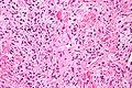
SFT - high mag. (WC/Nephron)
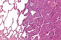
Synovial sarcoma - intermed. mag. (WC/Nephron)


Gross:
Differential diagnosis
Entities in which staghorn vessels are seen:
- Solitary fibrous tumour/hemangiopericytoma.[1]
- Malignant peripheral nerve sheath tumour (MPNST).[1]
- Synovial sarcoma.[1]
- Myofibroma.[1]
Others:[1]
- Mesenchymal chondrosarcoma.
- Infantile fibrosarcoma.
- Pleomorphic undifferentiated sarcoma.
- Leiomyosarcoma.
- Endometrial stromal sarcoma.
- Malignant mesothelioma.
- Thymoma
- Sarcomatoid carcinoma.
- Malignant melanoma.
- Glomus tumour.[2]
References
- ↑ 1.0 1.1 1.2 1.3 1.4 1.5 Nappi, O.; Ritter, JH.; Pettinato, G.; Wick, MR. (Aug 1995). "Hemangiopericytoma: histopathological pattern or clinicopathologic entity?". Semin Diagn Pathol 12 (3): 221-32. PMID 8545589.
- ↑ Slone, SP.; Moore, GD.; Parker, LP.; Rickard, KA.; Nixdorf-Miller, AS. (Jan 2010). "Glomus tumor of the ovary masquerading as granulosa cell tumor: case report.". Int J Gynecol Pathol 29 (1): 24-6. doi:10.1097/PGP.0b013e3181b0b771. PMID 19952942.