Bone
Jump to navigation
Jump to search
Bone is a scaffold it bears weight and occasionally gets infected.
Tumours often spread to bone and occasionally arise in bone. Bone tumours are dealt with in the bone tumours article.
Normal
- Normal bone has osteocytes.
- If the osteocytes are missing... the bone is dead.
- Osteoblasts - make bone.
- Osteoclasts - destroy bone.
Memory device: 'b' before 'c'.
Hyperostosis frontalis interna
- Extra-thick frontal bone.[1]
- No clinical significance -- just has to be recognized as a "nothing".
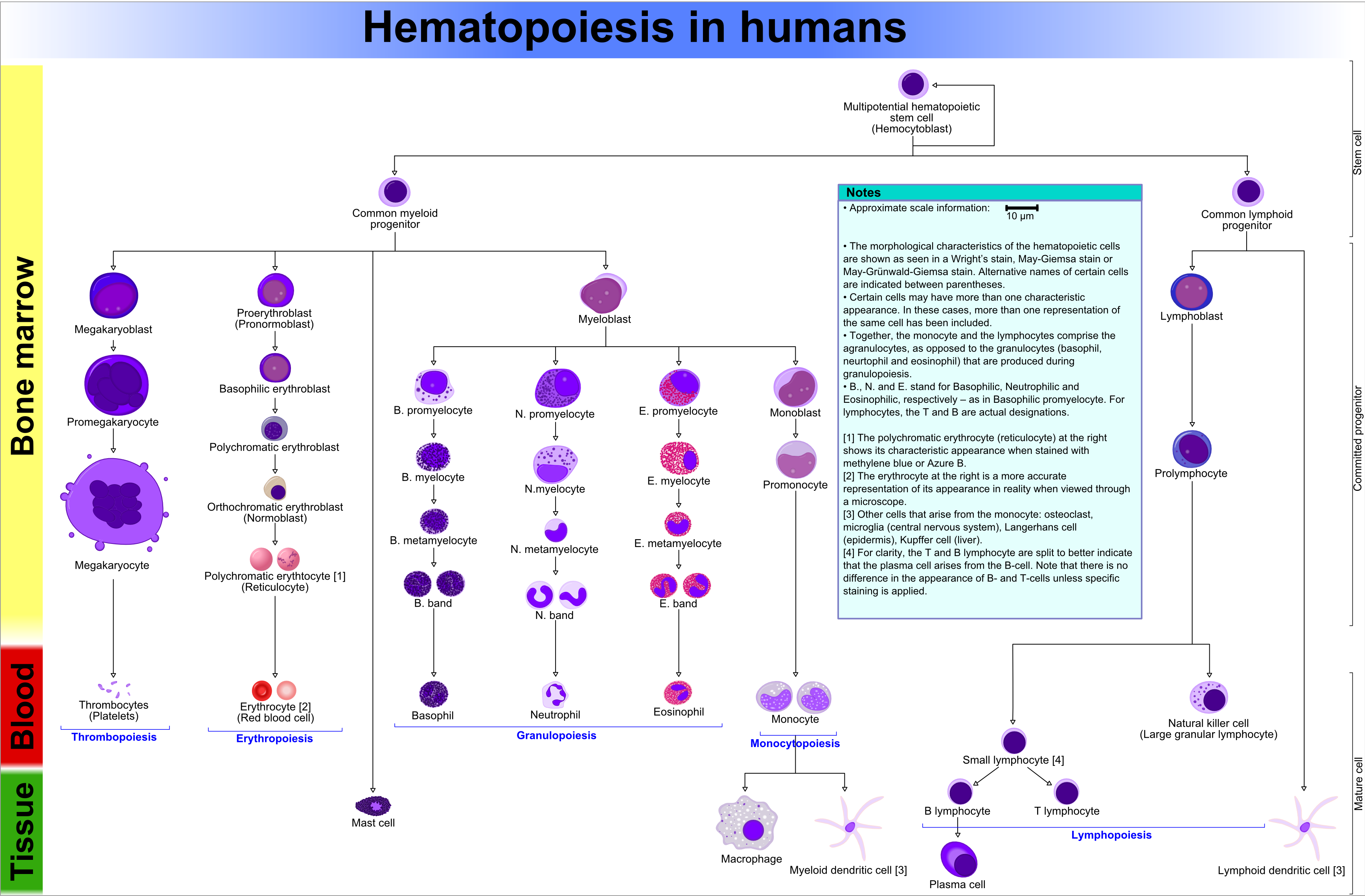
Bone marrow
Main article: Haematopoiesis
- Fat content (%) ~= age (in years)[2]
- e.g. 60 year old will have 60% fatty replacement.
- Should see three cell lines.
- The cell lines:[3]
- Erythroid (red cells),
- Myeloid (white blood cells),
- Megakaryocytic (platelets).
- The cell lines:[3]
Note: Lymphocytes are considered separately and typically spared in bone marrow failure.[4]
Identifying the lines:[5]
- Megakaryocytes:
- Big cells ~ 3x the size of a RBC.
- Normoblasts (RBC precursors):
- Hyperchromatic, i.e. blue, nucleus.
- Myeloid line:
- Granules.
- Reniform nucleus, i.e. kidney bean shaped nucleus.
Images:
Organization
- Mature hematopoeitic cells at the centre (distant from bone).
- Immature hematopoeitic cells adjacent to the bone.
Infectious
Osteomyelitis
General
- Hematogenous - often in children.
- Direct entry (skin defect) - adults with diabetes.
Microscopic
- PMNs.
Chronic osteomyelitis
- Plasma cells.
- May be sterile, i.e. no organisms.
Bone tumours
Main article: Bone tumours
This is a big topic. It is dealt with in a separate article.
The bone tumour article covers tumour mimics, e.g. brown cell tumour.
Fractures
Main article: Forensic pathology
This is dealt with in the forensic pathology article.
Others
The following is a collection of weird stuffs.
Myositis ossificans
Epidemiology:
- Young people.
- History of trauma.
Microscopic
Features:[6]
- High cellularity.
- Low mitotic activity.
- No atypical mitoses.
- No hyperchromasia.
Other features:[7]
- Low power diagnosis:
- Lesion is well-circumscribed.
- Normal muscle is adjacent to the lesion.
Paget disease of the bone
General
- Benign - unlike Paget disease of the breast.
Classically divided into three phases:[8][9]
- Lytic (predominantly osteoclasts).
- Mixed lytic (osteoclastic) and blastic (osteoblastic).
- Sclerotic (burned-out).
Clinical:
- Elevated ALP.
Microscopic
Features:[8]
- Bone matrix has jigsaw-puzzle like pattern.
- Jigsaw-puzzle pieces each ~ 100-500 micrometres in size (largest dimension).
- Increased osteoclast activity.
- Osteoclast = macrophage that resorbs bone matrix.
Images:
See also
References
- ↑ URL: http://radiopaedia.org/articles/hyperostosis_frontalis_interna. Accessed on: 29 September 2010.
- ↑ IAV. 26 Feb 2009.
- ↑ http://emedicine.medscape.com/article/199003-overview
- ↑ http://emedicine.medscape.com/article/199003-overview
- ↑ http://upload.wikimedia.org/wikipedia/commons/6/69/Hematopoiesis_%28human%29_diagram.png
- ↑ 6.0 6.1 Humphrey, Peter A; Dehner, Louis P; Pfeifer, John D (2008). The Washington Manual of Surgical Pathology (1st ed.). Lippincott Williams & Wilkins. pp. 607. ISBN 978-0781765275.
- ↑ IAV. 9 December 2010.
- ↑ 8.0 8.1 URL: http://emedicine.medscape.com/article/311688-overview. Accessed on: 25 December 2010.
- ↑ URL: http://radiopaedia.org/articles/paget-disease-of-bone-1. Accessed on: 25 December 2010.