Difference between revisions of "Chondrosarcoma"
Jump to navigation
Jump to search
(redirect) |
(split out) |
||
| Line 1: | Line 1: | ||
[[Chondrosarcoma]] is a malignant tumour of [[cartilage]]. It is in the [[Chondro-osseous_tumours|Chondro-osseous grouping of tumours]] and can be lumped into the much large category of the [[soft tissue lesions]]. | |||
==General== | |||
*Usually a good prognosis. | |||
Clinical/epidemiologic features:<ref name=pmid17976362>{{cite journal |author=Skubitz KM, D'Adamo DR |title=Sarcoma |journal=Mayo Clin. Proc. |volume=82 |issue=11 |pages=1409–32 |year=2007 |month=November |pmid=17976362 |doi= |url=http://www.mayoclinicproceedings.com/content/82/11/1409.long}}</ref> | |||
*Usually arise in a (benign) abnormality of cartilage (e.g. osteochondroma, enchondroma). | |||
*May be associated with a syndrome: | |||
**Olier disease (multiple enchondromatosis). | |||
**Maffucci syndrome (multiple enchondromas and hemangiomas). | |||
Notes: | |||
*Review article (from oncology perspective): PMID 17545802. | |||
===Subtypes=== | |||
Several subtypes exist: | |||
*Chondrosarcoma not otherwise specified (NOS). | |||
*Juxtacortical chondrosarcoma. | |||
*Myxoid chondrosarcoma. | |||
*Mesenchymal chondrosarcoma. | |||
*Clear cell chondrosarcoma. | |||
*Dedifferentiated chondrosarcoma. | |||
==Microscopic== | |||
Features:<ref>IAV. 26 February 2009.</ref><ref name=Ref_Klatt417>{{Ref Klatt|417}}</ref> | |||
*"Abnormal cartilage": | |||
**+/-Nuclear atypia - high grade lesions. | |||
***High grade lesions: | |||
****Nuclear clearing. | |||
****Nucleoli. | |||
****Hyperchromasia. | |||
***Low/intermediate grade lesions: | |||
****Bi-nucleation. | |||
****Hypochromatic enlarged nuclei. | |||
****Infiltration of lamellar bone ("invasion") - not common - '''diagnostic'''. | |||
**Increased cellularity. | |||
***More cellular than cartilage... but relatively paucicellular compared to other sarcomas. | |||
**Irregular spacing of chondrocytes. | |||
Notes: | |||
*Low grade chondrosarcoma are not cytologically malignant; the diagnosis rests mostly on radiologic findings. | |||
**The exception is ''infiltration of lamellar bone'' -- this is diagnostic of chondrosarcoma.<ref>Dickson, B. 28 April 2011.</ref> | |||
DDx: | |||
*[[Chordoma]]. | |||
*[[Enchondroma]]. | |||
*[[Synovial chondromatosis]]. | |||
*[[Osteosarcoma]] - esp. [[chondroblastic osteosarcoma]] - has osteoid, may be focal. | |||
====Images==== | |||
<gallery> | |||
Image:Chondrosarcoma_(1).jpg | Chondrosarcoma - low mag. (WC) | |||
Image:Chondrosarcoma_(2).jpg | Chondrosarcoma - high mag. (WC) | |||
Image:Chondrosarcoma_(3).jpg | Chondrosarcoma - high mag. (WC) | |||
</gallery> | |||
www: | |||
*[http://path.upmc.edu/cases/case168.html Chondrosarcoma (upmc.edu)]. | |||
*[http://www.path.utah.edu/casepath/ms%20cases/MSCase6/chondrosarcoma%20low%20grade%20sp03-9617%20g%20(Large)%20(Large).jpg Low-grade chondrosarcoma (path.utah.edu)].<ref>URL: [http://www.path.utah.edu/casepath/ms%20cases/MSCase6/MSCase6Part3.htm http://www.path.utah.edu/casepath/ms%20cases/MSCase6/MSCase6Part3.htm]. Accessed on: 29 December 2013.</ref> | |||
===Variants=== | |||
====Mesenchymal chondrosarcoma==== | |||
*Arise in soft tissue; this is where the name comes from.<ref name=pmid14161087>{{cite journal |author=Dowling EA |title=Mesenchymal chondrosarcoma |journal=J Bone Joint Surg Am |volume=46 |issue= |pages=747–54 |year=1964 |month=June |pmid=14161087 |doi= |url=http://www.ejbjs.org/cgi/reprint/46/4/747.pdf}}</ref> | |||
*Rare variant of chondrosarcoma. | |||
Microscopic: | |||
Features: | |||
*"White clouds in a blue sky". | |||
Image: | |||
*[http://moon.ouhsc.edu/kfung/jty1/opaq/PathQuiz/S0A001-PQ01-M.htm Mesenchymal chondrosarcoma (ouhsc.edu)]. | |||
====Myxoid chondrosarcoma==== | |||
Microscopic: | |||
Features: | |||
*[[Chordoma]]-like: | |||
**[[Myxoid]] background. | |||
**Small cells with eosinophilic cytoplasm. | |||
DDx: | |||
*Chondroid [[syringoma]]. | |||
*Parachordoma.<ref name=pmid10809219>{{cite journal |author=Fisher C |title=Parachordoma exists--but what is it? |journal=Adv Anat Pathol |volume=7 |issue=3 |pages=141–8 |year=2000 |month=May |pmid=10809219 |doi= |url=}}</ref> | |||
*[[Chordoma]]. (???) | |||
====Extraskeletal myxoid chondrosarcoma==== | |||
*Originally thought to be a variant of ''myxoid chondrosarcoma of bone''; however, may not be a chondrosarcoma at all.<ref name=pmid14657948>{{Cite journal | last1 = Aigner | first1 = T. | last2 = Oliveira | first2 = AM. | last3 = Nascimento | first3 = AG. | title = Extraskeletal myxoid chondrosarcomas do not show a chondrocytic phenotype. | journal = Mod Pathol | volume = 17 | issue = 2 | pages = 214-21 | month = Feb | year = 2004 | doi = 10.1038/modpathol.3800036 | PMID = 14657948 | URL = http://www.nature.com/modpathol/journal/v17/n2/full/3800036a.html }}</ref> | |||
*Characteristic [[chromosomal translocation]]: t(9;22) CHN-EWS. | |||
DDx: | |||
*Chordoma.<ref name=pmid14657948/> | |||
**S-100 +ve (strong). | |||
**EMA +ve. | |||
Image: | |||
*[http://www.cttr.org/large/03113.jpg Extraskeletal myxoid chondrosarcoma (cttr.org)].<ref>URL: [http://www.cttr.org/cms/?p=736 http://www.cttr.org/cms/?p=736]. Accessed on: 1 May 2011.</ref> | |||
====Dedifferentiated chondrosarcoma==== | |||
Clinical: | |||
*Abysmal to poor prognosis. | |||
**In one series (22 patients) 5-year survival ~20%.<ref>{{Cite journal | last1 = Mitchell | first1 = AD. | last2 = Ayoub | first2 = K. | last3 = Mangham | first3 = DC. | last4 = Grimer | first4 = RJ. | last5 = Carter | first5 = SR. | last6 = Tillman | first6 = RM. | title = Experience in the treatment of dedifferentiated chondrosarcoma. | journal = J Bone Joint Surg Br | volume = 82 | issue = 1 | pages = 55-61 | month = Jan | year = 2000 | doi = | PMID = 10697315 | URL = http://www.jbjs.org.uk/cgi/pmidlookup?view=long&pmid=10697315 }}</ref> | |||
**All dead in two years in another series (25 patients).<ref name=pmid17653766/> | |||
Features:<ref name=pmid17653766>{{Cite journal | last1 = Sopta | first1 = J. | last2 = Dordević | first2 = A. | last3 = Tulić | first3 = G. | last4 = Mijucić | first4 = V. | title = Dedifferentiated chondrosarcoma: our clinico-pathological experience and dilemmas in 25 cases. | journal = J Cancer Res Clin Oncol | volume = 134 | issue = 2 | pages = 147-52 | month = Feb | year = 2008 | doi = 10.1007/s00432-007-0262-5 | PMID = 17653766 }}</ref> | |||
#Poorly differentiated (mesenchymal) malignancy. | |||
#Well-differentiated cartilaginous component. | |||
Images: | |||
*[http://path.upmc.edu/cases/case118/micro.html Dedifferentiated chondrosarcoma (upmc.edu)]. | |||
===Grading=== | |||
Features:<ref name=Ref_WMSP643>{{Ref WMSP|643}}</ref> | |||
*Grade I: mild-to-moderate increase of cellularity +/- binucleated cells. | |||
*Grade II: between Grade I and Grade III. | |||
*Grade III: nuclear pleomorphism, mitoses common. | |||
==IHC== | |||
*S-100 -ve. (???) | |||
==See also== | |||
*[[Chondro-osseous tumours]]. | |||
==References== | |||
{{Reflist|2}} | |||
[[Category:Diagnosis]] | [[Category:Diagnosis]] | ||
[[Category:Chondro-osseous tumours]] | |||
Revision as of 02:33, 30 December 2013
Chondrosarcoma is a malignant tumour of cartilage. It is in the Chondro-osseous grouping of tumours and can be lumped into the much large category of the soft tissue lesions.
General
- Usually a good prognosis.
Clinical/epidemiologic features:[1]
- Usually arise in a (benign) abnormality of cartilage (e.g. osteochondroma, enchondroma).
- May be associated with a syndrome:
- Olier disease (multiple enchondromatosis).
- Maffucci syndrome (multiple enchondromas and hemangiomas).
Notes:
- Review article (from oncology perspective): PMID 17545802.
Subtypes
Several subtypes exist:
- Chondrosarcoma not otherwise specified (NOS).
- Juxtacortical chondrosarcoma.
- Myxoid chondrosarcoma.
- Mesenchymal chondrosarcoma.
- Clear cell chondrosarcoma.
- Dedifferentiated chondrosarcoma.
Microscopic
- "Abnormal cartilage":
- +/-Nuclear atypia - high grade lesions.
- High grade lesions:
- Nuclear clearing.
- Nucleoli.
- Hyperchromasia.
- Low/intermediate grade lesions:
- Bi-nucleation.
- Hypochromatic enlarged nuclei.
- Infiltration of lamellar bone ("invasion") - not common - diagnostic.
- High grade lesions:
- Increased cellularity.
- More cellular than cartilage... but relatively paucicellular compared to other sarcomas.
- Irregular spacing of chondrocytes.
- +/-Nuclear atypia - high grade lesions.
Notes:
- Low grade chondrosarcoma are not cytologically malignant; the diagnosis rests mostly on radiologic findings.
- The exception is infiltration of lamellar bone -- this is diagnostic of chondrosarcoma.[4]
DDx:
- Chordoma.
- Enchondroma.
- Synovial chondromatosis.
- Osteosarcoma - esp. chondroblastic osteosarcoma - has osteoid, may be focal.
Images
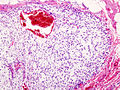
Chondrosarcoma - low mag. (WC)
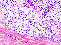
Chondrosarcoma - high mag. (WC)
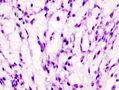
Chondrosarcoma - high mag. (WC)
.jpg)
.jpg)
.jpg)
www:
%20(Large).jpg)
Variants
Mesenchymal chondrosarcoma
- Arise in soft tissue; this is where the name comes from.[6]
- Rare variant of chondrosarcoma.
Microscopic: Features:
- "White clouds in a blue sky".
Image:
Myxoid chondrosarcoma
Microscopic: Features:
DDx:
Extraskeletal myxoid chondrosarcoma
- Originally thought to be a variant of myxoid chondrosarcoma of bone; however, may not be a chondrosarcoma at all.[8]
- Characteristic chromosomal translocation: t(9;22) CHN-EWS.
DDx:
- Chordoma.[8]
- S-100 +ve (strong).
- EMA +ve.
Image:
Dedifferentiated chondrosarcoma
Clinical:
- Abysmal to poor prognosis.
Features:[11]
- Poorly differentiated (mesenchymal) malignancy.
- Well-differentiated cartilaginous component.
Images:
Grading
Features:[12]
- Grade I: mild-to-moderate increase of cellularity +/- binucleated cells.
- Grade II: between Grade I and Grade III.
- Grade III: nuclear pleomorphism, mitoses common.
IHC
- S-100 -ve. (???)
See also
References
- ↑ Skubitz KM, D'Adamo DR (November 2007). "Sarcoma". Mayo Clin. Proc. 82 (11): 1409–32. PMID 17976362. http://www.mayoclinicproceedings.com/content/82/11/1409.long.
- ↑ IAV. 26 February 2009.
- ↑ Klatt, Edward C. (2006). Robbins and Cotran Atlas of Pathology (1st ed.). Saunders. pp. 417. ISBN 978-1416002741.
- ↑ Dickson, B. 28 April 2011.
- ↑ URL: http://www.path.utah.edu/casepath/ms%20cases/MSCase6/MSCase6Part3.htm. Accessed on: 29 December 2013.
- ↑ Dowling EA (June 1964). "Mesenchymal chondrosarcoma". J Bone Joint Surg Am 46: 747–54. PMID 14161087. http://www.ejbjs.org/cgi/reprint/46/4/747.pdf.
- ↑ Fisher C (May 2000). "Parachordoma exists--but what is it?". Adv Anat Pathol 7 (3): 141–8. PMID 10809219.
- ↑ 8.0 8.1 Aigner, T.; Oliveira, AM.; Nascimento, AG. (Feb 2004). "Extraskeletal myxoid chondrosarcomas do not show a chondrocytic phenotype.". Mod Pathol 17 (2): 214-21. doi:10.1038/modpathol.3800036. PMID 14657948.
- ↑ URL: http://www.cttr.org/cms/?p=736. Accessed on: 1 May 2011.
- ↑ Mitchell, AD.; Ayoub, K.; Mangham, DC.; Grimer, RJ.; Carter, SR.; Tillman, RM. (Jan 2000). "Experience in the treatment of dedifferentiated chondrosarcoma.". J Bone Joint Surg Br 82 (1): 55-61. PMID 10697315.
- ↑ 11.0 11.1 Sopta, J.; Dordević, A.; Tulić, G.; Mijucić, V. (Feb 2008). "Dedifferentiated chondrosarcoma: our clinico-pathological experience and dilemmas in 25 cases.". J Cancer Res Clin Oncol 134 (2): 147-52. doi:10.1007/s00432-007-0262-5. PMID 17653766.
- ↑ Humphrey, Peter A; Dehner, Louis P; Pfeifer, John D (2008). The Washington Manual of Surgical Pathology (1st ed.). Lippincott Williams & Wilkins. pp. 643. ISBN 978-0781765275.