Difference between revisions of "Vermiform appendix"
m (re-arrange, update) |
|||
| (108 intermediate revisions by 2 users not shown) | |||
| Line 1: | Line 1: | ||
The ''' | The '''vermiform appendix''', usually just '''appendix''', is a little thingy that is attached to the [[cecum]]. Taking it out is the bread 'n butter of [[general surgery]]. | ||
The appendix is a vestigial structure that is thought to have arisen from a larger cecum. Larger cecae are often seen in herbivores and thought to facilitate better digestion of plant matter.<ref>Dawkins R. The Greatest Show on Earth: The Evidence for Evolution | The appendix is a vestigial structure that is thought to have arisen from a larger cecum. Larger cecae are often seen in herbivores and thought to facilitate better digestion of plant matter.<ref>{{cite book |author=Dawkins, R. |title=The Greatest Show on Earth: The Evidence for Evolution |publisher=Free Press |location= |year=2009 |pages=115 |edition=1st |isbn=978-1416594789 |oclc= |doi= |accessdate=}}</ref> | ||
== | =Normal= | ||
==Normal vermiform appendix== | |||
===General=== | ===General=== | ||
* | *Seen in: | ||
**Right hemicolectomies. | |||
***[[colorectal carcinoma|Colon cancer]]. | |||
***[[Crohn's disease]]. | |||
**Surgeries for ovarian mucinous tumours. | |||
===Gross=== | ===Gross=== | ||
*Shiny serosal surface. | |||
* | **No exudate. | ||
* | *Normal diameter. | ||
*+/- | **6.6 +/- 1.5 mm -- based on CT.<ref name=pmid21344807>{{Cite journal | last1 = Charoensak | first1 = A. | last2 = Pongpornsup | first2 = S. | last3 = Suthikeeree | first3 = W. | title = Wall thickness and outer diameter of the normal appendix in adults using 64 slices multidetector CT. | journal = J Med Assoc Thai | volume = 93 | issue = 12 | pages = 1437-42 | month = Dec | year = 2010 | doi = | PMID = 21344807 }}</ref> | ||
=== | |||
===Microscopic=== | ===Microscopic=== | ||
Features: | Features: | ||
*+/-Lymphoid hyperplasia - mucosa or submucosa. | |||
* +/- | *Normal colorectal-type mucosa. | ||
* | *Fatty submucosa. | ||
*Benign smooth muscle. | |||
*Serosa. | |||
Negatives: | |||
* | *No [[neutrophil]]s in the muscularis propria. | ||
* | *No lesion in appendiceal tip. | ||
* | *No serosal inflammation ([[periappendicitis]]). | ||
* | *No organisms in the appendiceal lumen, e.g. [[Enterobius vermicularis]]. | ||
DDx: | |||
*[[Adenovirus appendicitis]]. | |||
* | *[[Cryptosporidiosis]]. | ||
*Mild colitis. | |||
===Sign out=== | |||
<pre> | |||
VERMIFORM APPENDIX WITHIN NORMAL LIMITS. | |||
</pre> | |||
Note: | |||
*This is for a normal appendix within a larger operation. The article ''[[negative appendectomy]]'' deals with a normal appearing appendix that was removed for presumed appendicitis. | |||
* | |||
== | ==Negative appendectomy== | ||
{{Main|Negative appendectomy}} | |||
An appendectomy done for presumed [[acute appendicitis]] that is pathologically within normal limits | |||
== | =Inflammatory pathologies= | ||
==Acute appendicitis== | |||
{{Main|Acute appendicitis}} | |||
==Adenovirus appendicitis== | |||
{{Main|Adenovirus appendicitis}} | |||
==Enterobius vermicularis== | |||
{| | {{Main|Enterobius vermicularis}} | ||
| | *[[AKA]] ''pinworm''. | ||
|- | ===General=== | ||
| | *May be found in the appendix. | ||
| | *The incidence is higher in normal appendices than inflamed ones.<ref name=pmid1853157>{{Cite journal | last1 = Wiebe | first1 = BM. | title = Appendicitis and Enterobius vermicularis. | journal = Scand J Gastroenterol | volume = 26 | issue = 3 | pages = 336-8 | month = Mar | year = 1991 | doi = | PMID = 1853157 }}</ref><ref name=pmid7945067/> | ||
| | *Clinically mimics appendicitis.<ref>{{cite journal |author=Ariyarathenam AV, Nachimuthu S, Tang TY, Courtney ED, Harris SA, Harris AM |title=Enterobius vermicularis infestation of the appendix and management at the time of laparoscopic appendectomy: case series and literature review |journal=Int J Surg |volume=8 |issue=6 |pages=466–9 |year=2010 |pmid=20637320 |doi=10.1016/j.ijsu.2010.06.007 |url=}}</ref> | ||
| | |||
| | |||
| | |||
| | |||
=== | ===Microscopic=== | ||
Features: | |||
* | *Usu. the appendiceal wall has no inflammation, i.e. there is no appendicitis.<ref name=pmid1853157/><ref name=pmid7945067>{{Cite journal | last1 = Dahlstrom | first1 = JE. | last2 = Macarthur | first2 = EB. | title = Enterobius vermicularis: a possible cause of symptoms resembling appendicitis. | journal = Aust N Z J Surg | volume = 64 | issue = 10 | pages = 692-4 | month = Oct | year = 1994 | doi = | PMID = 7945067 }}</ref> | ||
*''[[Enterobius vermicularis]]'' organisms. | |||
* | |||
====Image==== | |||
<gallery> | |||
Image:Enterobius_-_very_low_mag.jpg | Enterobius - very low mag. (WC/Nephron) | |||
Image:Enterobius_-_high_mag.jpg | Enterobius - high mag. (WC/Nephron) | |||
Image:Pinworms_in_the_Appendix_%281%29.jpg | Pinworm (WC/Uthman) | |||
</gallery> | |||
=== | ==Granulomatous appendicitis== | ||
{{Main|Granulomatous appendicitis}} | |||
=== | ==Inflammatory bowel disease== | ||
: See ''[[Inflammatory bowel disease]]''. | |||
== | ==Periappendicitis== | ||
===General=== | |||
Definition: inflammation of tissues around the (vermiform) appendix.<ref>URL: [http://www.medilexicon.com/medicaldictionary.php?t=66889 http://www.medilexicon.com/medicaldictionary.php?t=66889]. Accessed on: 1 June 2011.</ref> | |||
*May be seen in association of appendicitis or alone. | |||
**With appendicitis it is suggestive of perforation. | |||
**Without concurrent appendicitis it is suggestive of another abdominal pathology.<ref name=pmid2349982>{{Cite journal | last1 = Fink | first1 = AS. | last2 = Kosakowski | first2 = CA. | last3 = Hiatt | first3 = JR. | last4 = Cochran | first4 = AJ. | title = Periappendicitis is a significant clinical finding. | journal = Am J Surg | volume = 159 | issue = 6 | pages = 564-8 | month = Jun | year = 1990 | doi = | PMID = 2349982 }}</ref><ref>{{Cite journal | last1 = O'Neil | first1 = MB. | last2 = Moore | first2 = DB. | title = Periappendicitis: Clinical reality or pathologic curiosity? | journal = Am J Surg | volume = 134 | issue = 3 | pages = 356-7 | month = Sep | year = 1977 | doi = | PMID = 900337 }}</ref> | |||
=== | ===Microscopic=== | ||
* | Features: | ||
* | *Acute inflammation of the serosa. | ||
**[[Neutrophil]]s in the serosa. | |||
DDx: | |||
*[[Acute appendicitis]]. | |||
==== | =Tumours of the appendix= | ||
* | ==Adenocarcinoma== | ||
*Like ''colorectal adenocarcinoma'' - see ''[[colorectal tumours]]''. | |||
==Mucinous tumours of the appendix== | |||
{{Main|Mucinous tumours of the appendix}} | |||
This grouping includes ''mucinous cystadenoma'' and ''mucinous cystadenocarcinoma''. | |||
* | ==Goblet cell adenocarcinoma== | ||
{{Main|Goblet cell adenocarcinoma}} | |||
*Previously known as ''goblet cell carcinoid''. | |||
==Neuroendocrine tumour of the appendix== | |||
* | *Previously known as ''appendiceal carcinoid''. | ||
*[[AKA]] ''appendiceal neuroendocrine tumour'', abbreviated ''appendiceal NET''. | |||
{{Main|Neuroendocrine tumour of the appendix}} | |||
=See also= | |||
*[[Colon]]. | *[[Colon]]. | ||
*[[Gastrointestinal pathology]]. | *[[Gastrointestinal pathology]]. | ||
=References= | |||
{{reflist|2}} | {{reflist|2}} | ||
[[Category:Gastrointestinal pathology]] | [[Category:Gastrointestinal pathology]] | ||
[[Category:Vermiform appendix]] | |||
Latest revision as of 15:11, 4 December 2023
The vermiform appendix, usually just appendix, is a little thingy that is attached to the cecum. Taking it out is the bread 'n butter of general surgery.
The appendix is a vestigial structure that is thought to have arisen from a larger cecum. Larger cecae are often seen in herbivores and thought to facilitate better digestion of plant matter.[1]
Normal
Normal vermiform appendix
General
- Seen in:
- Right hemicolectomies.
- Surgeries for ovarian mucinous tumours.
Gross
- Shiny serosal surface.
- No exudate.
- Normal diameter.
- 6.6 +/- 1.5 mm -- based on CT.[2]
Microscopic
Features:
- +/-Lymphoid hyperplasia - mucosa or submucosa.
- Normal colorectal-type mucosa.
- Fatty submucosa.
- Benign smooth muscle.
- Serosa.
Negatives:
- No neutrophils in the muscularis propria.
- No lesion in appendiceal tip.
- No serosal inflammation (periappendicitis).
- No organisms in the appendiceal lumen, e.g. Enterobius vermicularis.
DDx:
- Adenovirus appendicitis.
- Cryptosporidiosis.
- Mild colitis.
Sign out
VERMIFORM APPENDIX WITHIN NORMAL LIMITS.
Note:
- This is for a normal appendix within a larger operation. The article negative appendectomy deals with a normal appearing appendix that was removed for presumed appendicitis.
Negative appendectomy
An appendectomy done for presumed acute appendicitis that is pathologically within normal limits
Inflammatory pathologies
Acute appendicitis
Adenovirus appendicitis
Enterobius vermicularis
- AKA pinworm.
General
- May be found in the appendix.
- The incidence is higher in normal appendices than inflamed ones.[3][4]
- Clinically mimics appendicitis.[5]
Microscopic
Features:
- Usu. the appendiceal wall has no inflammation, i.e. there is no appendicitis.[3][4]
- Enterobius vermicularis organisms.
Image
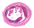
Enterobius - very low mag. (WC/Nephron)
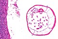
Enterobius - high mag. (WC/Nephron)
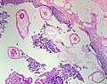
Pinworm (WC/Uthman)


.jpg)
Granulomatous appendicitis
Inflammatory bowel disease
Periappendicitis
General
Definition: inflammation of tissues around the (vermiform) appendix.[6]
- May be seen in association of appendicitis or alone.
Microscopic
Features:
- Acute inflammation of the serosa.
- Neutrophils in the serosa.
DDx:
Tumours of the appendix
Adenocarcinoma
- Like colorectal adenocarcinoma - see colorectal tumours.
Mucinous tumours of the appendix
This grouping includes mucinous cystadenoma and mucinous cystadenocarcinoma.
Goblet cell adenocarcinoma
- Previously known as goblet cell carcinoid.
Neuroendocrine tumour of the appendix
- Previously known as appendiceal carcinoid.
- AKA appendiceal neuroendocrine tumour, abbreviated appendiceal NET.
See also
References
- ↑ Dawkins, R. (2009). The Greatest Show on Earth: The Evidence for Evolution (1st ed.). Free Press. pp. 115. ISBN 978-1416594789.
- ↑ Charoensak, A.; Pongpornsup, S.; Suthikeeree, W. (Dec 2010). "Wall thickness and outer diameter of the normal appendix in adults using 64 slices multidetector CT.". J Med Assoc Thai 93 (12): 1437-42. PMID 21344807.
- ↑ 3.0 3.1 Wiebe, BM. (Mar 1991). "Appendicitis and Enterobius vermicularis.". Scand J Gastroenterol 26 (3): 336-8. PMID 1853157.
- ↑ 4.0 4.1 Dahlstrom, JE.; Macarthur, EB. (Oct 1994). "Enterobius vermicularis: a possible cause of symptoms resembling appendicitis.". Aust N Z J Surg 64 (10): 692-4. PMID 7945067.
- ↑ Ariyarathenam AV, Nachimuthu S, Tang TY, Courtney ED, Harris SA, Harris AM (2010). "Enterobius vermicularis infestation of the appendix and management at the time of laparoscopic appendectomy: case series and literature review". Int J Surg 8 (6): 466–9. doi:10.1016/j.ijsu.2010.06.007. PMID 20637320.
- ↑ URL: http://www.medilexicon.com/medicaldictionary.php?t=66889. Accessed on: 1 June 2011.
- ↑ Fink, AS.; Kosakowski, CA.; Hiatt, JR.; Cochran, AJ. (Jun 1990). "Periappendicitis is a significant clinical finding.". Am J Surg 159 (6): 564-8. PMID 2349982.
- ↑ O'Neil, MB.; Moore, DB. (Sep 1977). "Periappendicitis: Clinical reality or pathologic curiosity?". Am J Surg 134 (3): 356-7. PMID 900337.