Difference between revisions of "Sarcoidosis"
m (→See also: w) |
|||
| (33 intermediate revisions by 2 users not shown) | |||
| Line 1: | Line 1: | ||
'''Sarcoidosis''' is non-necrotizing granulomatous disease of unknown etiology. It classically associated with (pulmonary) hilar lymphadenopathy. It may be found in almost any organ | {{ Infobox diagnosis | ||
| Name = {{PAGENAME}} | |||
| Image = Sarcoidosis (1) lymph node biopsy.jpg | |||
| Width = | |||
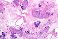
| Caption = Sarcoidosis-like granulomas in a lymph node. [[H&E stain]]. | |||
| Micro = well-formed [[granuloma]]s often with few surrounding lymphocytes ("naked"), usually non-necrotizing | |||
| Subtypes = | |||
| LMDDx = fungal infections, [[MAC]], [[tuberculosis]], other infections, drug reactions, reactive process to malignancy | |||
| Stains = AFB -ve, GMS -ve, PASD -ve | |||
| IHC = | |||
| EM = | |||
| Molecular = PCR for [[tuberculosis]] -ve | |||
| IF = | |||
| Gross = | |||
| Grossing = | |||
| Site = [[lung]], hilar lymph nodes of the lung, [[skin]], [[cardiac sarcoidosis|heart]], other sites | |||
| Assdx = | |||
| Syndromes = | |||
| Clinicalhx = | |||
| Signs = | |||
| Symptoms = | |||
| Prevalence = uncommon | |||
| Bloodwork = +/-ACE elevated | |||
| Rads = +/-bilateral hilar lymphadenopathy (very common), +/-interstitial pattern, +/- pulmonary infiltrates, +/-cystic/bullous changes | |||
| Endoscopy = | |||
| Prognosis = | |||
| Other = | |||
| ClinDDx = [[lymphoma]], [[metastatic]] carcinoma, [[Wegener's granulomatosis]], others | |||
}} | |||
'''Sarcoidosis''' is non-necrotizing [[granulomatous disease]] of unknown etiology. It classically associated with (pulmonary) hilar lymphadenopathy. It may be found in almost any organ. | |||
This article covers the topic in general and focuses on the [[lung]] aspects. [[Cardiac sarcoidosis]] is dealt with separately. | |||
==General== | ==General== | ||
*Diagnosis of exclusion - infection must be excluded. | *[[Diagnosis]] of exclusion - infection, neoplasm, and drugs must be excluded. | ||
* | *Uncommon. | ||
*Afflicits skin ~25% of the time.<ref name=pmid24138972>{{Cite journal | last1 = Noiles | first1 = K. | last2 = Beleznay | first2 = K. | last3 = Crawford | first3 = RI. | last4 = Au | first4 = S. | title = Sarcoidosis can present with necrotizing granulomas histologically: two cases of ulcerated sarcoidosis and review of the literature. | journal = J Cutan Med Surg | volume = 17 | issue = 6 | pages = 377-83 | month = | year = | doi = | PMID = 24138972 }}</ref> | |||
Serology: | |||
*Angiotensin-converting enzyme (ACE) - used for diagnosis and to monitor activity.<ref name=pmid24047001>{{Cite journal | last1 = Kaura | first1 = V. | last2 = Kaura | first2 = NV. | last3 = Kaura | first3 = BN. | last4 = Kaura | first4 = CS. | title = Angiotensin-converting enzyme inhibitors in the treatment of sarcoidosis and association with ACE gene polymorphism: case series. | journal = Indian J Chest Dis Allied Sci | volume = 55 | issue = 2 | pages = 105-7 | month = | year = | doi = | PMID = 24047001 }}</ref><ref name=pmid23897103>{{Cite journal | last1 = Stouten | first1 = K. | last2 = Werken | first2 = MV. | last3 = Tchetverikov | first3 = I. | last4 = Saboerali | first4 = M. | last5 = Vermeer | first5 = HJ. | last6 = Castel | first6 = R. | last7 = Verheijen | first7 = FM. | title = Extreme elevation of serum angiotensin-converting enzyme (ACE) activity: always consider familial ACE hyperactivity. | journal = Ann Clin Biochem | volume = | issue = | pages = | month = Jul | year = 2013 | doi = 10.1177/0004563213489812 | PMID = 23897103 }}</ref> | |||
**Elevated in approximately 65% of patients in one series.<ref name=pmid9327039>{{Cite journal | last1 = Shorr | first1 = AF. | last2 = Torrington | first2 = KG. | last3 = Parker | first3 = JM. | title = Serum angiotensin converting enzyme does not correlate with radiographic stage at initial diagnosis of sarcoidosis. | journal = Respir Med | volume = 91 | issue = 7 | pages = 399-401 | month = Aug | year = 1997 | doi = | PMID = 9327039 }}</ref> | |||
==Gross== | |||
*Lungs - classic location.<ref name=pmid23597964>{{Cite journal | last1 = Rao | first1 = DA. | last2 = Dellaripa | first2 = PF. | title = Extrapulmonary manifestations of sarcoidosis. | journal = Rheum Dis Clin North Am | volume = 39 | issue = 2 | pages = 277-97 | month = May | year = 2013 | doi = 10.1016/j.rdc.2013.02.007 | PMID = 23597964 }} | |||
</ref> | |||
*Bilateral hilar lymphadenopathy. | |||
DDx lungs - radiologic: | |||
*Carcinomatosis - interstitial pattern.<ref>URL: [http://www.radiologyassistant.nl/en/46b480a6e4bdc http://www.radiologyassistant.nl/en/46b480a6e4bdc]. Accessed on: 23 May 2010.</ref> | |||
*Lymphoma - bilateral lymphadenopathy +/- mediastinal lymphadenopathy.<ref name=pmid23207258>{{Cite journal | last1 = Boujaoude | first1 = Z. | last2 = Dahdel | first2 = M. | last3 = Pratter | first3 = M. | last4 = Kass | first4 = J. | title = Endobronchial ultrasound with transbronchial needle aspiration in the diagnosis of bilateral hilar and mediastinal lymphadenopathy. | journal = J Bronchology Interv Pulmonol | volume = 19 | issue = 1 | pages = 19-23 | month = Jan | year = 2012 | doi = 10.1097/LBR.0b013e3182442b89 | PMID = 23207258 }}</ref> | |||
<gallery> | |||
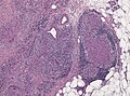
File:Sarcoidosis - Bilateral hilar lymphadenoathy (6076995153).jpg | Markedly enlarged bilaterally hilar lymph nodes in stage 1 sarcoidosis (WC/Yale Rosen) | |||
File:Sarcoidosis - Fibrosis (6076822244).jpg | Fibrosis (WC/Yale Rosen) | |||
</gallery> | |||
==Microscopic== | ==Microscopic== | ||
Features: | Features: | ||
*[[Granulomata]], well-formed, non-necrotizing. | *[[Granulomata]], well-formed, non-necrotizing. ‡ | ||
**Negative for microorganisms with special stains | **Negative for microorganisms with special stains. | ||
*Usu. minimal (lymphoid) inflammation; sarcoid granulomas are known as "naked granulomas".<ref name=pmid18948765>{{Cite journal | last1 = Brinster | first1 = NK. | title = Dermatopathology for the surgical pathologist: a pattern-based approach to the diagnosis of inflammatory skin disorders (part II). | journal = Adv Anat Pathol | volume = 15 | issue = 6 | pages = 350-69 | month = Nov | year = 2008 | doi = 10.1097/PAP.0b013e31818b1ac6 | PMID = 18948765 }}</ref> | *Usu. minimal (lymphoid) inflammation; sarcoid granulomas are known as "naked granulomas".<ref name=pmid18948765>{{Cite journal | last1 = Brinster | first1 = NK. | title = Dermatopathology for the surgical pathologist: a pattern-based approach to the diagnosis of inflammatory skin disorders (part II). | journal = Adv Anat Pathol | volume = 15 | issue = 6 | pages = 350-69 | month = Nov | year = 2008 | doi = 10.1097/PAP.0b013e31818b1ac6 | PMID = 18948765 }}</ref><ref name=pmid24138972>{{Cite journal | last1 = Noiles | first1 = K. | last2 = Beleznay | first2 = K. | last3 = Crawford | first3 = RI. | last4 = Au | first4 = S. | title = Sarcoidosis can present with necrotizing granulomas histologically: two cases of ulcerated sarcoidosis and review of the literature. | journal = J Cutan Med Surg | volume = 17 | issue = 6 | pages = 377-83 | month = | year = | doi = | PMID = 24138972 }}</ref> | ||
*In lung: | *In lung: | ||
**Interstitial location +/- centrilobular. | |||
Notes: | |||
*[[ | *‡ Reported with necrosis - uncommon.<ref name=pmid24138972>{{Cite journal | last1 = Noiles | first1 = K. | last2 = Beleznay | first2 = K. | last3 = Crawford | first3 = RI. | last4 = Au | first4 = S. | title = Sarcoidosis can present with necrotizing granulomas histologically: two cases of ulcerated sarcoidosis and review of the literature. | journal = J Cutan Med Surg | volume = 17 | issue = 6 | pages = 377-83 | month = | year = | doi = | PMID = 24138972 }}</ref> | ||
**[ | |||
DDx: | |||
*Infections. | |||
**Fungal. | |||
**Mycobacterial, e.g. [[tuberculosis]]. | |||
**Tertiary [[syphilis]].<ref name=pmid19249139>{{Cite journal | last1 = Hervier | first1 = B. | last2 = Wastiaux | first2 = H. | last3 = Freour | first3 = T. | last4 = Masseau | first4 = A. | last5 = Corvec | first5 = S. | last6 = Armingeat | first6 = T. | last7 = Hamidou | first7 = M. | title = [Sarcoidosis-like granulomatosis revealing a tertiary syphilis]. | journal = Rev Med Interne | volume = 30 | issue = 9 | pages = 806-8 | month = Sep | year = 2009 | doi = 10.1016/j.revmed.2009.01.003 | PMID = 19249139 }}</ref> | |||
*[[Hypersensitivity pneumonitis]] - usually poorly-formed granulomas, centrilobular, not paraseptal.{{fact}} | |||
*Reaction to treatment/drug. | |||
**Anti-TNF therapy.<ref name=pmid22389903>{{Cite journal | last1 = Tong | first1 = D. | last2 = Manolios | first2 = N. | last3 = Howe | first3 = G. | last4 = Spencer | first4 = D. | title = New onset sarcoid-like granulomatosis developing during anti-TNF therapy: an under-recognised complication. | journal = Intern Med J | volume = 42 | issue = 1 | pages = 89-94 | month = Jan | year = 2012 | doi = | PMID = 22389903 }}</ref> | |||
**Ipilimumab.<ref name=pmid24124863>{{Cite journal | last1 = Reule | first1 = RB. | last2 = North | first2 = JP. | title = Cutaneous and pulmonary sarcoidosis-like reaction associated with ipilimumab. | journal = J Am Acad Dermatol | volume = 69 | issue = 5 | pages = e272-3 | month = Nov | year = 2013 | doi = 10.1016/j.jaad.2013.07.028 | PMID = 24124863 }}</ref> | |||
*[[Common variable immunodeficiency]].<ref name=pmid18496983>{{Cite journal | last1 = Vultaggio | first1 = A. | last2 = Matucci | first2 = A. | last3 = Parronchi | first3 = P. | last4 = Rossi | first4 = O. | last5 = Filì | first5 = L. | last6 = Giudizi | first6 = MG. | last7 = Palandri | first7 = F. | last8 = Agostini | first8 = C. | last9 = Semenzato | first9 = G. | title = Association between sarcoidosis-like disease and common variable immunodeficiency (CVI): a new CVI variant showing an activation of the immune system. | journal = Sarcoidosis Vasc Diffuse Lung Dis | volume = 24 | issue = 2 | pages = 127-33 | month = Sep | year = 2007 | doi = | PMID = 18496983 }}</ref> | |||
*Reactive changes associated with tumours: | |||
**All testicular tumours<ref name=pmid18496978>{{Cite journal | last1 = Paparel | first1 = P. | last2 = Devonec | first2 = M. | last3 = Perrin | first3 = P. | last4 = Ruffion | first4 = A. | last5 = Decaussin-Petrucci | first5 = M. | last6 = Akin | first6 = O. | last7 = Sheinfeld | first7 = J. | last8 = Guillonneau | first8 = B. | title = Association between sarcoidosis and testicular carcinoma: a diagnostic pitfall. | journal = Sarcoidosis Vasc Diffuse Lung Dis | volume = 24 | issue = 2 | pages = 95-101 | month = Sep | year = 2007 | doi = | PMID = 18496978 }}</ref> - esp. [[seminoma]].<ref name=pmid17240627>{{Cite journal | last1 = Jankilevich | first1 = G. | last2 = Mendizabal | first2 = J. | last3 = Massa | first3 = MA. | last4 = Pedernera | first4 = A. | last5 = Galmes | first5 = M. | last6 = Spizzamiglio | first6 = N. | title = [Mediastinal sarcoidal reaction in follow up for seminoma]. | journal = Medicina (B Aires) | volume = 66 | issue = 6 | pages = 552-4 | month = | year = 2006 | doi = | PMID = 17240627 }}</ref> | |||
**[[Lung adenocarcinoma]].<ref name=pmid22499972>{{Cite journal | last1 = Tao | first1 = H. | last2 = Yamamoto | first2 = H. | last3 = Matsuda | first3 = E. | last4 = Sano | first4 = F. | last5 = Okabe | first5 = K. | last6 = Sugi | first6 = K. | title = Severe bronchoconstriction due to sarcoid-like reaction to lung cancer. | journal = Asian Cardiovasc Thorac Ann | volume = 20 | issue = 2 | pages = 199-201 | month = Apr | year = 2012 | doi = 10.1177/0218492311431493 | PMID = 22499972 }}</ref> | |||
===Images=== | |||
<gallery> | |||
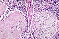
Image:Asteroid_body_intermed_mag.jpg | Sarcoidosis - lung. (WC) | |||
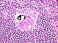
Image:Asteroid_body_very_high_mag.jpg | Granulomata in sarcoidosis with [[asteroid bodies]]. (WC) | |||
File:Sarcoidosis - Schaumann body (6151514639).jpg | [[Schaumann body]] in sarcoidosis. (WC/Yale Rosen) | |||
File:Sarcoidosis - Fibrosis of granulomas (6148634442).jpg | Fibrosis in sarcoid granulomas. (WC/Yale Rosen) | |||
File:Sarkoidosis muscle.jpg | Muscle involvement in sarcoidosis. (WC/jensflorian) | |||
File:Sarcoidosis_histology_skin_involvement.jpg | Skin involvement in sarcoidosis. (WC/jensflorian) | |||
</gallery> | |||
====Case==== | |||
<gallery> | |||
Image: Sarcoidosis - lung FNA -- very low mag.jpg | Sarcoidosis lung - very low mag. (WC) | |||
Image: Sarcoidosis - lung FNA -- low mag.jpg | Sarcoidosis lung - low mag. (WC) | |||
Image: Sarcoidosis - lung FNA -- intermed mag.jpg | Sarcoidosis lung - intermed. mag. (WC) | |||
Image: Sarcoidosis - lung FNA -- high mag.jpg | Sarcoidosis lung - high mag. (WC) | |||
Image: Sarcoidosis - lung FNA -- very high mag.jpg | Sarcoidosis lung - very high mag. (WC) | |||
</gallery> | |||
====www==== | |||
*[http://path.upmc.edu/cases/case412.html Sarcoidosis - several images (upmc.edu)]. | |||
*[http://path.upmc.edu/cases/case517.html Neurosarcoidosis - several images (upmc.edu)]. | |||
==Stains== | ==Stains== | ||
* | *[[AFB stain|AFB]] -ve. | ||
*PASD -ve. | *[[PASD stain|PASD]] -ve. | ||
*GMS -ve. | *[[GMS]] -ve. | ||
Note: | Note: | ||
* | *Must be done to exclude infection. | ||
==Molecular== | |||
*PCR for Tuberculosis -ve. | |||
==Sign out== | |||
*Should be something like ''sarcoid-like granulomas'' and ''clinical correlation required''. | |||
<pre> | |||
A. RIGHT LOWER LOBE LUNG, WEDGE RESECTION: | |||
- GRANULOMATOUS INFLAMMATION, NON-NECROTIZING (SARCOID-LIKE), SEE COMMENT. | |||
B. RIGHT MIDDLE LOBE LUNG, WEDGE RESECTION: | |||
- GRANULOMATOUS INFLAMMATION, NON-NECROTIZING (SARCOID-LIKE), SEE COMMENT. | |||
C. RIGHT LOWER LOBE LUNG, WEDGE RESECTION: | |||
- GRANULOMATOUS INFLAMMATION, NON-NECROTIZING (SARCOID-LIKE), SEE COMMENT. | |||
COMMENT: | |||
The sections show multiple sarcoid-like granulomas with both centrilobular and | |||
septal involvement. There is a slight upper lobe predominance of the disease. | |||
The main histomorphologic differential diagnoses are: sarcoidosis, | |||
infectious granulomatous pneumonia. | |||
Sarcoidosis is favoured based on the morphology of the granulomas and the | |||
lack of microorganisms with special stains (ZN, PASF, GMS). | |||
A serum ACE level should be considered, if not already done. | |||
Clinical and radiologic correlation is required. | |||
</pre> | |||
==See also== | ==See also== | ||
*[[Medical lung diseases]]. | *[[Medical lung diseases]]. | ||
*[[Cardiac sarcoidosis]]. | *[[Cardiac sarcoidosis]]. | ||
*[[Melkersson-Rosenthal syndrome]]. | |||
==References== | ==References== | ||
Latest revision as of 13:25, 18 October 2021
Sarcoidosis is non-necrotizing granulomatous disease of unknown etiology. It classically associated with (pulmonary) hilar lymphadenopathy. It may be found in almost any organ.
| Sarcoidosis | |
|---|---|
| Diagnosis in short | |
_lymph_node_biopsy.jpg) Sarcoidosis-like granulomas in a lymph node. H&E stain. | |
|
| |
| LM | well-formed granulomas often with few surrounding lymphocytes ("naked"), usually non-necrotizing |
| LM DDx | fungal infections, MAC, tuberculosis, other infections, drug reactions, reactive process to malignancy |
| Stains | AFB -ve, GMS -ve, PASD -ve |
| Molecular | PCR for tuberculosis -ve |
| Site | lung, hilar lymph nodes of the lung, skin, heart, other sites |
|
| |
| Prevalence | uncommon |
| Blood work | +/-ACE elevated |
| Radiology | +/-bilateral hilar lymphadenopathy (very common), +/-interstitial pattern, +/- pulmonary infiltrates, +/-cystic/bullous changes |
| Clin. DDx | lymphoma, metastatic carcinoma, Wegener's granulomatosis, others |
This article covers the topic in general and focuses on the lung aspects. Cardiac sarcoidosis is dealt with separately.
General
- Diagnosis of exclusion - infection, neoplasm, and drugs must be excluded.
- Uncommon.
- Afflicits skin ~25% of the time.[1]
Serology:
Gross
- Lungs - classic location.[5]
- Bilateral hilar lymphadenopathy.
DDx lungs - radiologic:
- Carcinomatosis - interstitial pattern.[6]
- Lymphoma - bilateral lymphadenopathy +/- mediastinal lymphadenopathy.[7]
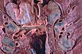
Markedly enlarged bilaterally hilar lymph nodes in stage 1 sarcoidosis (WC/Yale Rosen)
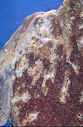
Fibrosis (WC/Yale Rosen)
.jpg)
.jpg)
Microscopic
Features:
- Granulomata, well-formed, non-necrotizing. ‡
- Negative for microorganisms with special stains.
- Usu. minimal (lymphoid) inflammation; sarcoid granulomas are known as "naked granulomas".[8][1]
- In lung:
- Interstitial location +/- centrilobular.
Notes:
- ‡ Reported with necrosis - uncommon.[1]
DDx:
- Infections.
- Fungal.
- Mycobacterial, e.g. tuberculosis.
- Tertiary syphilis.[9]
- Hypersensitivity pneumonitis - usually poorly-formed granulomas, centrilobular, not paraseptal.[citation needed]
- Reaction to treatment/drug.
- Common variable immunodeficiency.[12]
- Reactive changes associated with tumours:
- All testicular tumours[13] - esp. seminoma.[14]
- Lung adenocarcinoma.[15]
Images
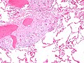
Sarcoidosis - lung. (WC)
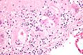
Granulomata in sarcoidosis with asteroid bodies. (WC)
Schaumann body in sarcoidosis. (WC/Yale Rosen)
Fibrosis in sarcoid granulomas. (WC/Yale Rosen)
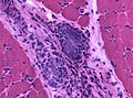
Muscle involvement in sarcoidosis. (WC/jensflorian)
Skin involvement in sarcoidosis. (WC/jensflorian)


.jpg)
.jpg)


Case
Sarcoidosis lung - very low mag. (WC)
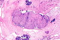
Sarcoidosis lung - low mag. (WC)
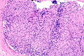
Sarcoidosis lung - intermed. mag. (WC)

Sarcoidosis lung - high mag. (WC)

Sarcoidosis lung - very high mag. (WC)


www
Stains
Note:
- Must be done to exclude infection.
Molecular
- PCR for Tuberculosis -ve.
Sign out
- Should be something like sarcoid-like granulomas and clinical correlation required.
A. RIGHT LOWER LOBE LUNG, WEDGE RESECTION:
- GRANULOMATOUS INFLAMMATION, NON-NECROTIZING (SARCOID-LIKE), SEE COMMENT.

B. RIGHT MIDDLE LOBE LUNG, WEDGE RESECTION:
- GRANULOMATOUS INFLAMMATION, NON-NECROTIZING (SARCOID-LIKE), SEE COMMENT.

C. RIGHT LOWER LOBE LUNG, WEDGE RESECTION:
- GRANULOMATOUS INFLAMMATION, NON-NECROTIZING (SARCOID-LIKE), SEE COMMENT.

COMMENT:
The sections show multiple sarcoid-like granulomas with both centrilobular and
septal involvement. There is a slight upper lobe predominance of the disease.

The main histomorphologic differential diagnoses are: sarcoidosis,
infectious granulomatous pneumonia.

Sarcoidosis is favoured based on the morphology of the granulomas and the
lack of microorganisms with special stains (ZN, PASF, GMS).

A serum ACE level should be considered, if not already done.

Clinical and radiologic correlation is required.
See also
References
- ↑ 1.0 1.1 1.2 Noiles, K.; Beleznay, K.; Crawford, RI.; Au, S.. "Sarcoidosis can present with necrotizing granulomas histologically: two cases of ulcerated sarcoidosis and review of the literature.". J Cutan Med Surg 17 (6): 377-83. PMID 24138972.
- ↑ Kaura, V.; Kaura, NV.; Kaura, BN.; Kaura, CS.. "Angiotensin-converting enzyme inhibitors in the treatment of sarcoidosis and association with ACE gene polymorphism: case series.". Indian J Chest Dis Allied Sci 55 (2): 105-7. PMID 24047001.
- ↑ Stouten, K.; Werken, MV.; Tchetverikov, I.; Saboerali, M.; Vermeer, HJ.; Castel, R.; Verheijen, FM. (Jul 2013). "Extreme elevation of serum angiotensin-converting enzyme (ACE) activity: always consider familial ACE hyperactivity.". Ann Clin Biochem. doi:10.1177/0004563213489812. PMID 23897103.
- ↑ Shorr, AF.; Torrington, KG.; Parker, JM. (Aug 1997). "Serum angiotensin converting enzyme does not correlate with radiographic stage at initial diagnosis of sarcoidosis.". Respir Med 91 (7): 399-401. PMID 9327039.
- ↑ Rao, DA.; Dellaripa, PF. (May 2013). "Extrapulmonary manifestations of sarcoidosis.". Rheum Dis Clin North Am 39 (2): 277-97. doi:10.1016/j.rdc.2013.02.007. PMID 23597964.
- ↑ URL: http://www.radiologyassistant.nl/en/46b480a6e4bdc. Accessed on: 23 May 2010.
- ↑ Boujaoude, Z.; Dahdel, M.; Pratter, M.; Kass, J. (Jan 2012). "Endobronchial ultrasound with transbronchial needle aspiration in the diagnosis of bilateral hilar and mediastinal lymphadenopathy.". J Bronchology Interv Pulmonol 19 (1): 19-23. doi:10.1097/LBR.0b013e3182442b89. PMID 23207258.
- ↑ Brinster, NK. (Nov 2008). "Dermatopathology for the surgical pathologist: a pattern-based approach to the diagnosis of inflammatory skin disorders (part II).". Adv Anat Pathol 15 (6): 350-69. doi:10.1097/PAP.0b013e31818b1ac6. PMID 18948765.
- ↑ Hervier, B.; Wastiaux, H.; Freour, T.; Masseau, A.; Corvec, S.; Armingeat, T.; Hamidou, M. (Sep 2009). "[Sarcoidosis-like granulomatosis revealing a tertiary syphilis].". Rev Med Interne 30 (9): 806-8. doi:10.1016/j.revmed.2009.01.003. PMID 19249139.
- ↑ Tong, D.; Manolios, N.; Howe, G.; Spencer, D. (Jan 2012). "New onset sarcoid-like granulomatosis developing during anti-TNF therapy: an under-recognised complication.". Intern Med J 42 (1): 89-94. PMID 22389903.
- ↑ Reule, RB.; North, JP. (Nov 2013). "Cutaneous and pulmonary sarcoidosis-like reaction associated with ipilimumab.". J Am Acad Dermatol 69 (5): e272-3. doi:10.1016/j.jaad.2013.07.028. PMID 24124863.
- ↑ Vultaggio, A.; Matucci, A.; Parronchi, P.; Rossi, O.; Filì, L.; Giudizi, MG.; Palandri, F.; Agostini, C. et al. (Sep 2007). "Association between sarcoidosis-like disease and common variable immunodeficiency (CVI): a new CVI variant showing an activation of the immune system.". Sarcoidosis Vasc Diffuse Lung Dis 24 (2): 127-33. PMID 18496983.
- ↑ Paparel, P.; Devonec, M.; Perrin, P.; Ruffion, A.; Decaussin-Petrucci, M.; Akin, O.; Sheinfeld, J.; Guillonneau, B. (Sep 2007). "Association between sarcoidosis and testicular carcinoma: a diagnostic pitfall.". Sarcoidosis Vasc Diffuse Lung Dis 24 (2): 95-101. PMID 18496978.
- ↑ Jankilevich, G.; Mendizabal, J.; Massa, MA.; Pedernera, A.; Galmes, M.; Spizzamiglio, N. (2006). "[Mediastinal sarcoidal reaction in follow up for seminoma].". Medicina (B Aires) 66 (6): 552-4. PMID 17240627.
- ↑ Tao, H.; Yamamoto, H.; Matsuda, E.; Sano, F.; Okabe, K.; Sugi, K. (Apr 2012). "Severe bronchoconstriction due to sarcoid-like reaction to lung cancer.". Asian Cardiovasc Thorac Ann 20 (2): 199-201. doi:10.1177/0218492311431493. PMID 22499972.