Small cell carcinoma of the urinary bladder
(Redirected from Small cell carcinoma of the bladder)
Jump to navigation
Jump to search
| Small cell carcinoma of the urinary bladder | |
|---|---|
| Diagnosis in short | |
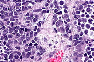
 Small cell carcinoma of the urinary bladder. H&E stain. | |
|
| |
| LM | small cohesive cells with scant cytoplasm, nuclear moulding |
| LM DDx | small cell carcinoma of the prostate gland, lymphoma (large cell), other small round blue cell tumours, urothelial carcinoma with a small cell carcinoma component |
| Stains | pankeratin +ve, chromogranin +ve, synaptophysin +ve, CD56 +ve |
| Site | urinary bladder |
|
| |
| Symptoms | +/-hematuria |
| Prevalence | very rare |
| Prognosis | poor |
| Clin. DDx | other bladder tumours esp. urothelial carcinoma |
Small cell carcinoma of the urinary bladder, abbreviated SCCUB, is a rare malignant neoplasm of the urinary bladder.
General
- Very rare[1] - less than 1% of bladder cancers in one series.[2]
- Poor prognosis[1] - survival ~ 11 months.[2]
Clinical:
- Hematuria - typical presentation.[2]
Epidemiology:
Microscopic
Features:
- Cohesive small cells (~2x resting lymphocytes) with scant cytoplasm - see small cell carcinoma.
- +/-Nuclear moulding.
- If mixed histology with urothelial carcinoma, small cell carcinoma component must be majority of tumour; otherwise classified as urothelial carcinoma with small cell carcinoma component.[3]
Note:
- Usually co-exists with urothelial carcinoma ~60% of cases in one series.[2]
DDx:
- Urothelial carcinoma with a small cell carcinoma component
- Small cell carcinoma of the prostate gland.
- Lymphoma, large cell.
- Other small round blue cell tumours.
Images
IHC
- Pankeratin +ve,
- Chromogranin +ve.
- Synaptophysin +ve.
- CD56 +ve.
- TTF-1 +ve/-ve.
Sign out
Urinary Bladder Tumour, Transurethral Resection:
     - SMALL CELL CARCINOMA, invades muscularis propria, see comment.
     -- Please see synoptic report.

Comment:
The tumour consists of cohesive, small blue cells with nuclear moulding and necrosis.  There is no definite urothelial differentiation, and no other differentiation to suggest a particular primary site.

The tumour stains as follows:
POSITIVE: CK7 (dot-like moderate, diffuse), chromogranin A (moderate, diffuse), synaptophysin (strong, diffuse), TTF-1 (strong, diffuse).
NEGATIVE: CK20, CD45 (lymphocytes in background), CD20, PSA, p63 (marks benign urothelium).
PROLIFERATION (Ki-67): >90%.

Small cell carcinoma of the urinary bladder is the main consideration, based the anatomical site of the specimen. The findings above should be considered in the context of the imaging and clinical history.
See also
References
- ↑ 1.0 1.1 Koga, F.; Yokoyama, M.; Fukushima, H. (Nov 2013). "Small cell carcinoma of the urinary bladder: a contemporary review with a special focus on bladder-sparing treatments.". Expert Rev Anticancer Ther 13 (11): 1269-79. doi:10.1586/14737140.2013.851605. PMID 24168010.
- ↑ 2.0 2.1 2.2 2.3 2.4 Hou, CP.; Lin, YH.; Chen, CL.; Chang, PL.; Tsui, KH. (2013). "Clinical outcome of primary small cell carcinoma of the urinary bladder.". Onco Targets Ther 6: 1179-85. doi:10.2147/OTT.S49879. PMID 24009428.
- ↑ The International Agency for Research on Cancer (Author), H. Moch (Editor), P.A. Humphrey (Editor), T.M. Ulbright (Editor), V.E. Reuter (Editor) (2016). WHO Classification of Tumours of the Urinary System and Male Genital Organs (4th ed.). Lyon: World Health Organization. pp. 117-8. ISBN 978-9283224372.


