Partial colectomy for diverticular disease
Jump to navigation
Jump to search
.jpg)
Partial colectomy for diverticular disease is a very common procedure. The sigmoid colon is typically afflicited; in that case it can more precisely be labeled sigmoidectomy for diverticular disease.
Introduction
This is a relatively common specimen. Diverticulitis (inflammation of diverticula) and it complications are usually diagnosed by computed tomography (CT).[1]
If the individual has a peritonitis, a (temporary) stoma is created in a surgery known as a Hartmann's procedure.[1]
The pathologist's main tasks in this specimen is:
- Confirming and documenting extent of the disease.
- Excluding malignancy.
Protocol
Specimen:
- Length __ cm.
- Circumference (proximal/one end) __ cm.
- Circumference (distal/other end) __ cm.
- Mesentry (maximal): __ cm.
Appearance:
- Serosal surface: [shiny/hemorrhagic/dull/exudate/adhesions].
- Mucosa: [unremarkable/granular].
- Polyps: [none/number - size __ cm, location (to nearest resection margin): __ cm].
- Number of diverticula (count up to 6, then estimate): [number of diverticula].
- Wall: [unremarkable/thickened].
Other:
- Perforation: [not identified/present - location of perforation (to nearest mucosal margin): __ cm, size of performation __ cm].
Representative sections submitted:
- Proximal mucosal margin.
- Distal mucosal margin.
- Diverticula (3-5 blocks).
- Interdiverticular mucosa (2 blocks).
- Lymph nodes (1 or 2 blocks). ‡
Protocol notes
‡ The Royal College of Pathologists (UK) recommends submitting lymph nodes in benign resection.[2]
Alternate approaches
Pathology
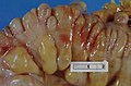
Diverticular disease.

See also
Related protocols
References
- ↑ 1.0 1.1 Schultz, JK.; Yaqub, S.; Øresland, T. (Oct 2016). "Management of Diverticular Disease in Scandinavia.". J Clin Gastroenterol 50 Suppl 1: S50-2. doi:10.1097/MCG.0000000000000642. PMID 27622365.
- ↑ URL: https://www.rcpath.org/static/4593f557-d75c-4ca6-9307a9d688e02a2d/g085-tp-giandp-jan16.pdf. Accessed on: 2024 Oct 15.