Acquired cystic disease-associated renal cell carcinoma
Jump to navigation
Jump to search
| Acquired cystic disease-associated renal cell carcinoma | |
|---|---|
| Diagnosis in short | |
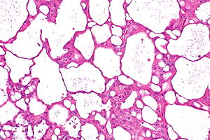
 Acquired cystic disease-associated renal cell carcinoma. H&E stain. | |
|
| |
| LM | sieve-like architecture (tubular structures/cribriforming), tumour cells with prominent nucleoli and eosinophilic cytoplasm, oxalate crystals - seen best in polarized light, acquired cystic disease in background, +/-papillary structures - common minor component |
| LM DDx | acquired cystic renal disease, papillary renal cell carcinoma, hereditary leiomyomatosis renal cell carcinoma syndrome associated renal cell carcinoma |
| IHC | AMACR +ve, CD10 +ve, pankeratin +ve, CK7 +ve (heterogeneous), EMA -ve |
| Gross | kidney with cystic changes, thinned cortex |
| Grossing notes | total nephrectomy for tumour grossing, partial nephrectomy grossing |
| Staging | kidney cancer staging |
| Site | kidney - see kidney tumours |
|
| |
| Associated Dx | acquired cystic disease of the kidney, end-stage renal disease |
| Prevalence | rare |
| Radiology | renal mass and cysts |
| Treatment | nephrectomy |
Acquired cystic disease-associated renal cell carcinoma, abbreviated ACD-RCC, is a rare kidney cancer that arises in the context of chronic renal failure.
It was added to the WHO classification of renal neoplasia in the Vancouver modification of 2012/2013.[1]
General
- Arise in the context of acquired cystic disease of the kidney which is seen in the context of end-stage renal disease (ESRD).[2]
- May have aggressive course - including lymph node metastases and death.[3]
Gross
- Cysts.
Microscopic
Features:
- Fused tubular structures/cribriforming/sieve-like architecture.[4]
- Focal papillary architecture - common.[3]
- Tumour cells have prominent nucleoli (ISUP nucleolar grade 3) and eosinophilic cytoplasm.
- Oxalate crystals - important.
- Look somewhat like cholesterol clefts.
- Seen easily in polarized light.
- Acquired cystic disease in background - required.
- Changes of end-stage kidney (obsolete glomeruli, thyroidization, interstitial fibrosis).
DDx:
- Acquired cystic disease of the kidney.
- Papillary renal cell carcinoma.
- Hereditary leiomyomatosis renal cell carcinoma syndrome associated renal cell carcinoma.
Images
Case
www
IHC
Features:[2]
- AMACR +ve.
- CD10 +ve.
- Pankeratin +ve.
- CK7 +ve (heterogeneous).
Others:[2]
See also
References
- ↑ Srigley, JR.; Delahunt, B.; Eble, JN.; Egevad, L.; Epstein, JI.; Grignon, D.; Hes, O.; Moch, H. et al. (Oct 2013). "The International Society of Urological Pathology (ISUP) Vancouver Classification of Renal Neoplasia.". Am J Surg Pathol 37 (10): 1469-89. doi:10.1097/PAS.0b013e318299f2d1. PMID 24025519.
- ↑ 2.0 2.1 2.2 Ahn, S.; Kwon, GY.; Cho, YM.; Jun, SY.; Choi, C.; Kim, HJ.; Park, YW.; Park, WS. et al. (Mar 2013). "Acquired cystic disease-associated renal cell carcinoma: further characterization of the morphologic and immunopathologic features.". Med Mol Morphol. doi:10.1007/s00795-013-0028-x. PMID 23471757.
- ↑ 3.0 3.1 Tickoo, SK.; dePeralta-Venturina, MN.; Harik, LR.; Worcester, HD.; Salama, ME.; Young, AN.; Moch, H.; Amin, MB. (Feb 2006). "Spectrum of epithelial neoplasms in end-stage renal disease: an experience from 66 tumor-bearing kidneys with emphasis on histologic patterns distinct from those in sporadic adult renal neoplasia.". Am J Surg Pathol 30 (2): 141-53. PMID 16434887.
- ↑ Srigley, JR.; Delahunt, B.; Eble, JN.; Egevad, L.; Epstein, JI.; Grignon, D.; Hes, O.; Moch, H. et al. (Oct 2013). "The International Society of Urological Pathology (ISUP) Vancouver Classification of Renal Neoplasia.". Am J Surg Pathol 37 (10): 1469-89. doi:10.1097/PAS.0b013e318299f2d1. PMID 24025519.
- ↑ 5.0 5.1 URL: https://www.auanet.org/education/modules/pathology/kidney-carcinomas/acquired-cystic.cfm. Accessed on: 13 May 2015.
- ↑ 6.0 6.1 6.2 6.3 Amin, Mahul B.; Eble, John; Grignon, David; Srigley, John. (2013). Urological Pathology (1st ed.). Wolters Kluwer. pp. 113. ISBN 978-0781782814.


