Difference between revisions of "Xanthogranulomatous pyelonephritis"
Jump to navigation
Jump to search
(+cat.) |
(split out) |
||
| Line 1: | Line 1: | ||
'''Xanthogranulomatous pyelonephritis''', abbreviated '''XGP''', is an inflammatory process of the [[kidney]] that can mimic [[renal cell carcinoma]]. | |||
==General== | |||
*May mimic [[renal cell carcinoma]] - especially radiologically. | |||
*Usually lower pole.{{fact}} | |||
*Associated with: | |||
**[[Diabetes mellitus]]. | |||
**History of UTI.<ref name=pmid17987581>{{cite journal |author=Afgan F, Mumtaz S, Ather MH |title=Preoperative diagnosis of xanthogranulomatous pyelonephritis |journal=Urol J |volume=4 |issue=3 |pages=169–73 |year=2007 |pmid=17987581 |doi= |url=}}</ref> | |||
**Nephrolithiasis. | |||
**GU obstruction.<ref name=pmid17098659>{{cite journal |author=Al-Ghazo MA, Ghalayini IF, Matalka II, Al-Kaisi NS, Khader YS |title=Xanthogranulomatous pyelonephritis: Analysis of 18 cases |journal=Asian J Surg |volume=29 |issue=4 |pages=257–61 |year=2006 |month=October |pmid=17098659 |doi= |url=}}</ref> | |||
*Occasionally an indication of nephrectomy.<ref name=pmid17987581/><ref name=pmid17098659/> | |||
*Most common organism (in the context of nephrectomy specimens) - ''Proteus mirabilis''.<ref name=pmid17098659/> | |||
==Microscopic== | |||
*Abundant macrophages. | |||
*+/-Giant cells. | |||
DDx: | |||
*[[Malakoplakia]]. | |||
**Basophilic inclusions -- inside or outside of macrophages - often size of RBC or larger ([[Michaelis-Gutmann bodies]]). | |||
*RCC - especially [[PRCC]] (as this has foamy macrophages). | |||
*Granulomatous disease. | |||
*[[Chronic pyelonephritis]]. | |||
*[[Interstitial nephritis]]. | |||
===Image=== | |||
<gallery> | |||
Image:Xanthogranulomatous_pyelonephritis_cd68.jpg | Xanthogranulomatous pyelonephritis - CD68 stain. (WC/Nephron) | |||
</gallery> | |||
==Stains== | |||
*[[PAS-D]] -ve. | |||
**Done to look for malakoplakia. | |||
==IHC== | |||
*CD68 +ve. | |||
*RCC markers (CD10, RCC) all negative. | |||
==Sign out== | |||
<pre> | |||
RIGHT KIDNEY, NEPHRECTOMY: | |||
- XANTHOGRANULOMATOUS PYELONEPHRITIS. | |||
- CHRONIC INTERSTITIAL NEPHRITIS. | |||
- INCREASED NUMBERS OF TOTALLY SCLEROSED GLOMERULI AND GLOMERULI WITH FOCAL | |||
SEGMENTAL GLOMERULOSCLEROSIS. | |||
- MARKED INTERSTITIAL FIBROSIS. | |||
- NEGATIVE FOR MALIGNANCY. | |||
COMMENT: | |||
Immunostaining demonstrates abundant CD68 positive cells (macrophages). A CD10 immunostain | |||
is non-concerning (it highlights glomeruli). A pankeratin immunostain is non-concerning | |||
(it highlights benign renal tubules). | |||
</pre> | |||
===Compatible XGP=== | |||
<pre> | |||
"KIDNEY" LESION, LEFT, BIOPSY: | |||
- FIBROMUSCULAR TISSUE WITH A MIXED INFLAMMATORY INFILTRATE. | |||
- CELLULAR DEBRIS WITH SURROUNDING LOOSELY AGGREGATED HISTIOCYTES. | |||
- NO RENAL PARENCHYMA IDENTIFIED. | |||
- NEGATIVE FOR MALIGNANCY. | |||
COMMENT: | |||
A SMA immunostain highlights the muscle component, and a CD68 immunostain marks | |||
abundant histiocytes. No epithelial component is identified with a pankeratin | |||
immunostain. | |||
</pre> | |||
==See also== | |||
*[[Kidney tumours]]. | |||
==References== | |||
{{Reflist|2}} | |||
[[Category:Diagnosis]] | [[Category:Diagnosis]] | ||
[[Category:Kidney tumours]] | |||
Revision as of 12:30, 5 April 2014
Xanthogranulomatous pyelonephritis, abbreviated XGP, is an inflammatory process of the kidney that can mimic renal cell carcinoma.
General
- May mimic renal cell carcinoma - especially radiologically.
- Usually lower pole.[citation needed]
- Associated with:
- Diabetes mellitus.
- History of UTI.[1]
- Nephrolithiasis.
- GU obstruction.[2]
- Occasionally an indication of nephrectomy.[1][2]
- Most common organism (in the context of nephrectomy specimens) - Proteus mirabilis.[2]
Microscopic
- Abundant macrophages.
- +/-Giant cells.
DDx:
- Malakoplakia.
- Basophilic inclusions -- inside or outside of macrophages - often size of RBC or larger (Michaelis-Gutmann bodies).
- RCC - especially PRCC (as this has foamy macrophages).
- Granulomatous disease.
- Chronic pyelonephritis.
- Interstitial nephritis.
Image
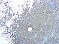
Xanthogranulomatous pyelonephritis - CD68 stain. (WC/Nephron)

Stains
- PAS-D -ve.
- Done to look for malakoplakia.
IHC
- CD68 +ve.
- RCC markers (CD10, RCC) all negative.
Sign out
RIGHT KIDNEY, NEPHRECTOMY:
- XANTHOGRANULOMATOUS PYELONEPHRITIS.
- CHRONIC INTERSTITIAL NEPHRITIS.
- INCREASED NUMBERS OF TOTALLY SCLEROSED GLOMERULI AND GLOMERULI WITH FOCAL
  SEGMENTAL GLOMERULOSCLEROSIS.
- MARKED INTERSTITIAL FIBROSIS.
- NEGATIVE FOR MALIGNANCY.

COMMENT:
Immunostaining demonstrates abundant CD68 positive cells (macrophages).  A CD10 immunostain
is non-concerning (it highlights glomeruli). A pankeratin immunostain is non-concerning
(it highlights benign renal tubules).
Compatible XGP
"KIDNEY" LESION, LEFT, BIOPSY:
- FIBROMUSCULAR TISSUE WITH A MIXED INFLAMMATORY INFILTRATE.
- CELLULAR DEBRIS WITH SURROUNDING LOOSELY AGGREGATED HISTIOCYTES.
- NO RENAL PARENCHYMA IDENTIFIED.
- NEGATIVE FOR MALIGNANCY.

COMMENT:
A SMA immunostain highlights the muscle component, and a CD68 immunostain marks
abundant histiocytes. No epithelial component is identified with a pankeratin
immunostain.
See also
References
- ↑ 1.0 1.1 Afgan F, Mumtaz S, Ather MH (2007). "Preoperative diagnosis of xanthogranulomatous pyelonephritis". Urol J 4 (3): 169–73. PMID 17987581.
- ↑ 2.0 2.1 2.2 Al-Ghazo MA, Ghalayini IF, Matalka II, Al-Kaisi NS, Khader YS (October 2006). "Xanthogranulomatous pyelonephritis: Analysis of 18 cases". Asian J Surg 29 (4): 257–61. PMID 17098659.