Viruses
Jump to navigation
Jump to search
This article collects all things virus. The more general topic of infective things is dealt with in microorganisms.
Many virus afflict humans. Only a few of them can be diagnosed histologically.
Viral inclusions - types
Cowdry types:[1]
- Cowdry type A inclusion:[2]
- Round eosinophilic material surrounded by a clear halo.
- Cowdry type B inclusion:[3]
- Neuropathology thingy. (???)
Images:
Viruses
Herpes simplex virus
- Abbreviated HSV.
General
Several subtypes:
- Canker sores - usually HSV-1.
- Genital herpes - usually HSV-2.
Histology/cytology
Features:[4]
- Clear "ground glass" nuclei.
- Rim of peripheral chromatin.
- Nuclear inclusions.
- Multinucleation with nuclear molding, i.e. multiple nuclei that touch over a large surface area.
Mnemonic - 3 Ms: Margination, Multinucleation, Molding.
Images:
- Herpes simplex virus - multinucleation (virology.org).
- HSV on a Pap test - showing multinucleation (WC).
- HSV esophagitis - very high mag. (WC).
- HSV esophagitis - intermed. mag. (WC).
Cytomegalovirus
- Abbreviated CMV
General
- The name comes from the microscopic appearance.
Microscopic
Features:
- Very large nucleus (as the name implies) with clearing.
- Granular cytoplasmic inclusions (red on H&E sections).
Notes:
- Classically in endothelial cells.
- In the context of esophageal ulcers, it is therefore useful to biopsy the base of the ulcer - if this is suspected.
Images:
IHC
- IHC for CMV is available - highlights granular cytoplasmic inclusions; increases sensitivity.
Human papilloma virus
- Abbreviated HPV.
General
- Causes a lot of pathology:
- Benign:
- Malignant:
- Cervical cancer and precursors (LSIL, HSIL).
- Anal cancer and precurors (AIN)
- Squamous cell carcinoma in the head & neck.
Oncocytic types:
Recombinant vaccine (Gardasil, Silgard) - covers:[6]
- HPV 6.
- HPV 11.
- HPV 16.
- HPV 18.
Microscopic
Features:
- Koilocytes:
- Perinuclear clearing.
- Nuclear changes.
- Size similar (or larger) to those in the basal layer of the epithelium.
- Nuclear enlargement should be evident on low power, i.e. 25x.
- Central location - nucleus should be smack in the middle of the cell.
Images:
IHC
- p16 +ve -- stains most cells infected by HPV.
Adenovirus
General
- Common in kids.
- May be seen in the context of (adenovirus) appendicitis.
Microscopic
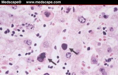
Features:
- "Smudge" cells[7] - black/blue blob ~ 10-15 micrometers. (???)
Notes:
Images:
- Adenovirus (medscape.com).[8]
- Smudge cell (medpedia.com).
- Necrosis in germinal centre - low mag. (flickr.com).
- Viral inclusions - high mag. (flickr.com).
- IHC for adenovirus (flickr.com)
Parvovirus
- AKA Parvovirus B19.
General
- Most significant in pregnant women.
- Parvovirus attacks the nucleated RBCs of the fetus - causes an aplastic anemia.
Trivia:
Microscopic
Features:
- Glassy (red) nuclear inclusions.[11]
- Nuclear enlargement.
Images:
- WC:
- www:
Epstein-Barr virus
- Abbreviated EBV
General
- Cases mononucleosis.
- Part of the herpes group of viruses.[14]
Associations
Cancer:[15]
- Classical Hodgkin lymphoma.
- Burkitt lymphoma.
- Nasopharyngeal carcinoma.
- B cell lymphomas -- in immunosuppressed individuals.
Microscopic
Features:
- Variable - see specific pathologies in Associations section.
- +/-Atypical lymphocytes.
- +/-Hemophagocytosis (uncommon).
Polyomavirus
May refer to:
- BK virus.
- Merkel cell polyomavirus.
- See: Merkel cell carcinoma.
See also
References
- ↑ URL: http://www.pathconsultddx.com/pathCon/largeImage?pii=S1559-8675%2806%2970864-6&figureId=fig3&ecomponentId=mmc3. Accessed: 12 January 2010.
- ↑ URL: http://www.whonamedit.com/synd.cfm/3495.html. Accessed on: 22 January 2010.
- ↑ http://www.whonamedit.com/synd.cfm/3496.html. Accessed on: 22 January 2010.
- ↑ SM. 11 January 2010.
- ↑ 5.0 5.1 De Boer, MA.; Peters, LA.; Aziz, MF.; Siregar, B.; Cornain, S.; Vrede, MA.; Jordanova, ES.; Fleuren, GJ. (Apr 2005). "Human papillomavirus type 18 variants: histopathology and E6/E7 polymorphisms in three countries.". Int J Cancer 114 (3): 422-5. doi:10.1002/ijc.20727. PMID 15551313.
- ↑ McCormack, PL.; Joura, EA. (Oct 2011). "Spotlight on Quadrivalent Human Papillomavirus(Types 6, 11, 16, 18) Recombinant Vaccine(Gardasil®) in the Prevention of PremalignantGenital Lesions, Genital Cancer, and Genital Warts in Women†.". BioDrugs 25 (5): 339-43. doi:10.2165/11205060-000000000-00000. PMID 21942919.
- ↑ URL: http://www.pathguy.com/lectures/infect.htm. Accessed on: 8 July 2010.
- ↑ URL:http://www.medscape.com/viewarticle/438534_2. Accessed on: 8 July 2010.
- ↑ Cossart, YE.; Field, AM.; Cant, B.; Widdows, D. (Jan 1975). "Parvovirus-like particles in human sera.". Lancet 1 (7898): 72-3. PMID 46024.
- ↑ Servey JT, Reamy BV, Hodge J (February 2007). "Clinical presentations of parvovirus B19 infection". Am Fam Physician 75 (3): 373–6. PMID 17304869. http://www.aafp.org/afp/991001ap/1455.html.
- ↑ URL: http://www.pathguy.com/lectures/infect.htm. Accessed on: 8 July 2010.
- ↑ URL:http://info.fujita-hu.ac.jp/~tsutsumi/case/case210.htm. Accessed on: 8 February 2011.
- ↑ URL: http://www.scielo.br/scielo.php?pid=S0036-46652007000200007&script=sci_arttext. Accessed on: 18 August 2011.
- ↑ URL: http://www.meddean.luc.edu/lumen/MedEd/orfpath/herpes.htm. Accessed on: 14 April 2011.
- ↑ Mitchell, Richard; Kumar, Vinay; Fausto, Nelson; Abbas, Abul K.; Aster, Jon (2011). Pocket Companion to Robbins & Cotran Pathologic Basis of Disease (8th ed.). Elsevier Saunders. pp. 169. ISBN 978-1416054542.
- ↑ Jin YK, Xie ZD, Yang S, Lu G, Shen KL (June 2010). "Epstein-Barr virus-associated hemophagocytic lymphohistiocytosis: a retrospective study of 78 pediatric cases in mainland of China". Chin. Med. J. 123 (11): 1426–30. PMID 20819601.