Difference between revisions of "Vascular tumours"
Jump to navigation
Jump to search
m (→IHC: +fli-1) |
|||
| (77 intermediate revisions by the same user not shown) | |||
| Line 1: | Line 1: | ||
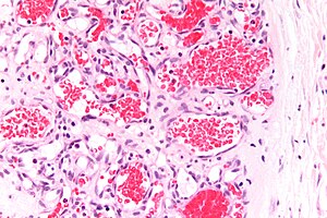
This article covers [[soft tissue lesions|soft tissue]] '''vascular | [[Image:Capillary_hemangioma_-_very_high_mag.jpg|thumb|right|[[Micrograph]] showing a [[capillary hemangioma]], a common type of vascular tumour. [[H&E stain]].]] | ||
This article covers [[soft tissue lesions|soft tissue]] '''vascular tumours'''. Vascular malformations are covered in the ''[[vascular malformations]]'' article. | |||
=Normal histology= | |||
[[Vascular_disease#Normal_blood_vessels|Normal blood vessel histology]] is dealt with in the ''[[vascular disease]]'' article. | [[Vascular_disease#Normal_blood_vessels|Normal blood vessel histology]] is dealt with in the ''[[vascular disease]]'' article. | ||
=Mimics= | |||
*[[Vascular malformations]]. | |||
**[[Arteriovenous malformation]]. | |||
**[[Varix]]. | |||
**[[Venous angioma]]. | |||
**[[Caverous malformation]]. | |||
*[[Angiomatoid fibrous histiocytoma]]. | |||
=Distinct entities= | |||
==Hemangioma== | ==Hemangioma== | ||
{{Main|Hemangioma}} | |||
{{Main|Liver hemangioma}} | |||
=== | ==Lymphangioma== | ||
*[[AKA]] ''cystic hygroma''.<ref name=Ref_WMSP12>{{Ref WMSP|12}}</ref> | |||
* | |||
===General=== | ===General=== | ||
* | *Benign. | ||
* | *Classically in the left neck.<ref name=Ref_WMSP12>{{Ref WMSP|12}}</ref> | ||
* | *May be seen in [[Turner syndrome]]. | ||
Treatment: | |||
*Surgical excision. | |||
===Microscopic=== | ===Microscopic=== | ||
Features:<ref name= | Features:<ref name=Ref_WMSP489>{{Ref WMSP|489}}</ref><ref name=Ref_WMSP12>{{Ref WMSP|12}}</ref> | ||
*Thin-walled channels lined by endothelium. | |||
*+/-Eosinophilic intraluminal material. | |||
*+/- | *+/-Clusters of intraluminal lymphocytes. | ||
*+/-Occasional [[RBC]]s. | |||
DDx: | DDx: | ||
* | *[[Hemangioma]]. | ||
Images: | Images: | ||
*[http:// | *[http://www.webpathology.com/image.asp?n=5&Case=412 Lymphangioma - low mag. (webpathology.com)]. | ||
*[http:// | *[http://www.webpathology.com/image.asp?case=412&n=6 Lymphangioma - high mag. (webpathology.com)]. | ||
===IHC=== | ===IHC=== | ||
Features:<ref name=pmid19098468>{{Cite journal | last1 = Kalof | first1 = AN. | last2 = Cooper | first2 = K. | title = D2-40 immunohistochemistry--so far! | journal = Adv Anat Pathol | volume = 16 | issue = 1 | pages = 62-4 | month = Jan | year = 2009 | doi = 10.1097/PAP.0b013e3181915e94 | PMID = 19098468 }}</ref><ref name=pmid11950918>{{Cite journal | last1 = Kahn | first1 = HJ. | last2 = Bailey | first2 = D. | last3 = Marks | first3 = A. | title = Monoclonal antibody D2-40, a new marker of lymphatic endothelium, reacts with Kaposi's sarcoma and a subset of angiosarcomas. | journal = Mod Pathol | volume = 15 | issue = 4 | pages = 434-40 | month = Apr | year = 2002 | doi = 10.1038/modpathol.3880543 | PMID = 11950918 }}</ref> | |||
*D2-40 +ve. | |||
* | |||
==Kaposi sarcoma== | |||
{{Main|Kaposi sarcoma}} | |||
==Masson hemangioma== | ==Masson hemangioma== | ||
*[[AKA]] ''intravascular papillary endothelial hyperplasia''.<ref name=pmid15865098>{{cite journal |author=Korkolis DP, Papaevangelou M, Koulaxouzidis G, Zirganos N, Psichogiou H, Vassilopoulos PP |title=Intravascular papillary endothelial hyperplasia (Masson's hemangioma) presenting as a soft-tissue sarcoma |journal=Anticancer Res. |volume=25 |issue=2B |pages=1409–12 |year=2005 |pmid=15865098 |doi= |url=}}</ref> | *[[AKA]] ''intravascular papillary endothelial hyperplasia''.<ref name=pmid15865098>{{cite journal |author=Korkolis DP, Papaevangelou M, Koulaxouzidis G, Zirganos N, Psichogiou H, Vassilopoulos PP |title=Intravascular papillary endothelial hyperplasia (Masson's hemangioma) presenting as a soft-tissue sarcoma |journal=Anticancer Res. |volume=25 |issue=2B |pages=1409–12 |year=2005 |pmid=15865098 |doi= |url=}}</ref> | ||
* | *[[AKA]] ''Masson tumor''.<ref>URL: [http://path.upmc.edu/cases/case544/dx.html http://path.upmc.edu/cases/case544/dx.html]. Accessed on: 25 January 2012.</ref> | ||
{{Main|intravascular papillary endothelial hyperplasia}} | |||
==Angiosarcoma== | ==Angiosarcoma== | ||
{{Main|Angiosarcoma}} | |||
==Kaposiform hemangioendothelioma== | ==Kaposiform hemangioendothelioma== | ||
===General=== | ===General=== | ||
*Locally aggressive.<ref name=Ref_WMSP603>{{Ref WMSP|603}}</ref> | *Locally aggressive.<ref name=Ref_WMSP603>{{Ref WMSP|603}}</ref> | ||
*Childhood tumour.<ref name=pmid15105642>{{Cite journal | last1 = Lyons | first1 = LL. | last2 = North | first2 = PE. | last3 = Mac-Moune Lai | first3 = F. | last4 = Stoler | first4 = MH. | last5 = Folpe | first5 = AL. | last6 = Weiss | first6 = SW. | title = Kaposiform hemangioendothelioma: a study of 33 cases emphasizing its pathologic, immunophenotypic, and biologic uniqueness from juvenile hemangioma. | journal = Am J Surg Pathol | volume = 28 | issue = 5 | pages = 559-68 | month = May | year = 2004 | doi = | PMID = 15105642 }}</ref> | |||
*Approximately half have ''Kasabach–Merritt phenomenon''<ref name=pmid15105642/> = vascular tumour --> coagulopathy. | |||
===Microscopic=== | ===Microscopic=== | ||
Features: | Features:<ref name=pmid1706068/> | ||
* | *Spindle cells lesions in sheets or nodules. | ||
*+/-Round tumour nodules - "cannon ball" appearance. | |||
DDx: | |||
*[[Kaposi sarcoma]].<ref name=pmid1706068>{{Cite journal | last1 = Miller | first1 = K. | title = Sister-chromatid exchange in human B- and T-lymphocytes exposed to bleomycin, cyclophosphamide, and ethyl methanesulfonate. | journal = Mutat Res | volume = 247 | issue = 1 | pages = 175-82 | month = Mar | year = 1991 | doi = | PMID = 1706068 | url = http://www.nature.com/modpathol/journal/v14/n11/full/3880441a.html }}</ref> | |||
===IHC=== | ===IHC=== | ||
Features:<ref name=pmid1706068/> | |||
*Vimentin +ve. | |||
*C31 +ve. | |||
*CD34 +ve. | |||
*UEA-1 lectin +ve. | |||
==Epithelioid hemangioendothelioma== | ==Epithelioid hemangioendothelioma== | ||
{{Main|Epithelioid hemangioendothelioma}} | |||
=== | ==Retiform hemangioendothelioma== | ||
{{Main|Retiform hemangioendothelioma}} | |||
==Intimal sarcoma== | |||
{{Main|Intimal sarcoma}} | |||
=See also= | |||
*[[Cardiovascular pathology]]. | *[[Cardiovascular pathology]]. | ||
*[[Vasculitides]]. | *[[Vasculitides]]. | ||
*[[Soft tissue lesions]]. | *[[Soft tissue lesions]]. | ||
=References= | |||
{{Reflist|2}} | {{Reflist|2}} | ||
[[Category:Vascular tumours]] | |||
[[Category:Soft tissue lesions]] | [[Category:Soft tissue lesions]] | ||
Latest revision as of 17:15, 18 January 2024

This article covers soft tissue vascular tumours. Vascular malformations are covered in the vascular malformations article.
Normal histology
Normal blood vessel histology is dealt with in the vascular disease article.
Mimics
Distinct entities
Hemangioma
Main article: Hemangioma
Main article: Liver hemangioma
Lymphangioma
General
- Benign.
- Classically in the left neck.[1]
- May be seen in Turner syndrome.
Treatment:
- Surgical excision.
Microscopic
- Thin-walled channels lined by endothelium.
- +/-Eosinophilic intraluminal material.
- +/-Clusters of intraluminal lymphocytes.
- +/-Occasional RBCs.
DDx:
Images:
IHC
- D2-40 +ve.
Kaposi sarcoma
Main article: Kaposi sarcoma
Masson hemangioma
Main article: intravascular papillary endothelial hyperplasia
Angiosarcoma
Main article: Angiosarcoma
Kaposiform hemangioendothelioma
General
- Locally aggressive.[7]
- Childhood tumour.[8]
- Approximately half have Kasabach–Merritt phenomenon[8] = vascular tumour --> coagulopathy.
Microscopic
Features:[9]
- Spindle cells lesions in sheets or nodules.
- +/-Round tumour nodules - "cannon ball" appearance.
DDx:
IHC
Features:[9]
- Vimentin +ve.
- C31 +ve.
- CD34 +ve.
- UEA-1 lectin +ve.
Epithelioid hemangioendothelioma
Main article: Epithelioid hemangioendothelioma
Retiform hemangioendothelioma
Main article: Retiform hemangioendothelioma
Intimal sarcoma
Main article: Intimal sarcoma
See also
References
- ↑ 1.0 1.1 1.2 Humphrey, Peter A; Dehner, Louis P; Pfeifer, John D (2008). The Washington Manual of Surgical Pathology (1st ed.). Lippincott Williams & Wilkins. pp. 12. ISBN 978-0781765275.
- ↑ Humphrey, Peter A; Dehner, Louis P; Pfeifer, John D (2008). The Washington Manual of Surgical Pathology (1st ed.). Lippincott Williams & Wilkins. pp. 489. ISBN 978-0781765275.
- ↑ Kalof, AN.; Cooper, K. (Jan 2009). "D2-40 immunohistochemistry--so far!". Adv Anat Pathol 16 (1): 62-4. doi:10.1097/PAP.0b013e3181915e94. PMID 19098468.
- ↑ Kahn, HJ.; Bailey, D.; Marks, A. (Apr 2002). "Monoclonal antibody D2-40, a new marker of lymphatic endothelium, reacts with Kaposi's sarcoma and a subset of angiosarcomas.". Mod Pathol 15 (4): 434-40. doi:10.1038/modpathol.3880543. PMID 11950918.
- ↑ Korkolis DP, Papaevangelou M, Koulaxouzidis G, Zirganos N, Psichogiou H, Vassilopoulos PP (2005). "Intravascular papillary endothelial hyperplasia (Masson's hemangioma) presenting as a soft-tissue sarcoma". Anticancer Res. 25 (2B): 1409–12. PMID 15865098.
- ↑ URL: http://path.upmc.edu/cases/case544/dx.html. Accessed on: 25 January 2012.
- ↑ Humphrey, Peter A; Dehner, Louis P; Pfeifer, John D (2008). The Washington Manual of Surgical Pathology (1st ed.). Lippincott Williams & Wilkins. pp. 603. ISBN 978-0781765275.
- ↑ 8.0 8.1 Lyons, LL.; North, PE.; Mac-Moune Lai, F.; Stoler, MH.; Folpe, AL.; Weiss, SW. (May 2004). "Kaposiform hemangioendothelioma: a study of 33 cases emphasizing its pathologic, immunophenotypic, and biologic uniqueness from juvenile hemangioma.". Am J Surg Pathol 28 (5): 559-68. PMID 15105642.
- ↑ 9.0 9.1 9.2 Miller, K. (Mar 1991). "Sister-chromatid exchange in human B- and T-lymphocytes exposed to bleomycin, cyclophosphamide, and ethyl methanesulfonate.". Mutat Res 247 (1): 175-82. PMID 1706068. http://www.nature.com/modpathol/journal/v14/n11/full/3880441a.html.