Ureter
Jump to navigation
Jump to search
The ureter is the tubule that takes the urine from the kidney to the urinary bladder. It is uncommonly afflicted by pathology that the pathologist sees on a day-to-day basis.
Normal ureter
Microscopic
Features:[1]
- Mucosa - epithelium and fibrous tissue.
- Muscularis:
- Upper 2/3: inner longitudinal, outer circular.
- Lower 1/3: inner longitudinal, middle circular, outer longitudinal.
Note:
- There is no submucosa!
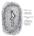
Ureter - cross-section. (WC)

Pathology of the ureter - overview
- Kidney stones.
- Ureteritis cystica.
- Urothelial neoplasias, esp. in Lynch syndrome[2] - see urothelium.
- Malakoplakia.
- Others.
Specific conditions
Ureteritis cystica
General
- Similar cystitis cystica.
- Uncommon.
- Painful.[3]
- Related to von Brunn's nests.[4]
Gross
- Smooth/round projections into the lumen.
Images:
Microscopic
Features:
- Nests of urothelium within the lamina propria with cyst formation, i.e. lumens are present.
Image:
Urothelial carcinoma
General
- Should prompt consideration of Lynch syndrome.
Microscopic
See:
See also
References
- ↑ URL: http://www.histology.leeds.ac.uk/urinary/ureter.php. Accessed: 31 October 2013.
- ↑ Crockett, DG.; Wagner, DG.; Holmäng, S.; Johansson, SL.; Lynch, HT. (May 2011). "Upper urinary tract carcinoma in Lynch syndrome cases.". J Urol 185 (5): 1627-30. doi:10.1016/j.juro.2010.12.102. PMID 21419447.
- ↑ Padilla-Fernández, B.; Díaz-Alférez, F.; Herrero-Polo, M.; Martín-Izquierdo, M.; Silva-Abuín, J.; Lorenzo-Gómez, M. (2012). "Ureteritis cystica: important consideration in the differential diagnosis of acute renal colic.". Clin Med Insights Case Rep 5: 29-33. doi:10.4137/CCRep.S9189. PMID 22474406.
- ↑ 4.0 4.1 4.2 Rothschild, JG.; Wu, G. (2011). "Ureteritis cystica: a radiologic pathologic correlation.". J Clin Imaging Sci 1: 23. doi:10.4103/2156-7514.80375. PMC 3177432. PMID 21966620. https://www.ncbi.nlm.nih.gov/pmc/articles/PMC3177432/.