Difference between revisions of "Ureter"
Jump to navigation
Jump to search
(fix sp.) |
|||
| (12 intermediate revisions by the same user not shown) | |||
| Line 1: | Line 1: | ||
The '''ureter''' is uncommonly afflicted by pathology that the pathologist sees on a day-to-day basis. | The '''ureter''' is the tubule that takes the urine from the [[kidney]] to the [[urinary bladder]]. It is uncommonly afflicted by pathology that the pathologist sees on a day-to-day basis. | ||
=Normal ureter= | |||
===Microscopic=== | |||
Features:<ref>URL: [http://www.histology.leeds.ac.uk/urinary/ureter.php http://www.histology.leeds.ac.uk/urinary/ureter.php]. Accessed: 31 October 2013.</ref> | |||
*Mucosa - epithelium and fibrous tissue. | |||
*Muscularis: | |||
**Upper 2/3: inner longitudinal, outer circular. | |||
**Lower 1/3: inner longitudinal, middle circular, outer longitudinal. | |||
Note: | |||
*There is no submucosa! | |||
<gallery> | |||
Image:Gray1134.png| Ureter - cross-section. (WC) | |||
</gallery> | |||
=Pathology of the ureter - overview= | |||
*[[Kidney stones]]. | |||
*[[Ureteritis cystica]]. | |||
*[[Urothelial carcinoma in situ]] - strong association with CIS in the urinary bladder.<ref name=pmid25374918>{{Cite journal | last1 = Zhou | first1 = H. | last2 = Ro | first2 = JY. | last3 = Truong | first3 = LD. | last4 = Ayala | first4 = AG. | last5 = Shen | first5 = SS. | title = Intraoperative frozen section evaluation of ureteral and urethral margins: studies of 203 consecutive radical cystoprostatectomy for men with bladder urothelial carcinoma. | journal = Am J Clin Exp Urol | volume = 2 | issue = 2 | pages = 156-60 | month = | year = 2014 | doi = | PMID = 25374918 }}</ref> | |||
*Other urothelial neoplasias - esp. in [[Lynch syndrome]]<ref name=pmid21419447>{{Cite journal | last1 = Crockett | first1 = DG. | last2 = Wagner | first2 = DG. | last3 = Holmäng | first3 = S. | last4 = Johansson | first4 = SL. | last5 = Lynch | first5 = HT. | title = Upper urinary tract carcinoma in Lynch syndrome cases. | journal = J Urol | volume = 185 | issue = 5 | pages = 1627-30 | month = May | year = 2011 | doi = 10.1016/j.juro.2010.12.102 | PMID = 21419447 }}</ref> - see [[urothelium]]. | |||
*[[Malakoplakia]]. | |||
*Others. | |||
=Specific conditions= | |||
==Ureteritis cystica== | ==Ureteritis cystica== | ||
===General=== | ===General=== | ||
| Line 11: | Line 35: | ||
*Smooth/round projections into the lumen. | *Smooth/round projections into the lumen. | ||
Images: | |||
*[http://www.ncbi.nlm.nih.gov/pmc/articles/PMC3177432/figure/F1/ Endoscopic image of ureteritis cystica (nih.gov)].<ref name=pmid21966620>{{Cite journal | last1 = Rothschild | first1 = JG. | last2 = Wu | first2 = G. | title = Ureteritis cystica: a radiologic pathologic correlation. | journal = J Clin Imaging Sci | volume = 1 | issue = | pages = 23 | month = | year = 2011 | doi = 10.4103/2156-7514.80375 | PMID = 21966620 | PMC = 3177432 | URL = http://www.ncbi.nlm.nih.gov/pmc/articles/PMC3177432/?tool=pubmed }}</ref> | *[http://www.ncbi.nlm.nih.gov/pmc/articles/PMC3177432/figure/F1/ Endoscopic image of ureteritis cystica (nih.gov)].<ref name=pmid21966620>{{Cite journal | last1 = Rothschild | first1 = JG. | last2 = Wu | first2 = G. | title = Ureteritis cystica: a radiologic pathologic correlation. | journal = J Clin Imaging Sci | volume = 1 | issue = | pages = 23 | month = | year = 2011 | doi = 10.4103/2156-7514.80375 | PMID = 21966620 | PMC = 3177432 | URL = http://www.ncbi.nlm.nih.gov/pmc/articles/PMC3177432/?tool=pubmed }}</ref> | ||
*[http://www.webpathology.com/image.asp?case=50&n=11 Ureteritis cystica (webpathology.com)]. | |||
===Microscopic=== | ===Microscopic=== | ||
| Line 21: | Line 46: | ||
*[http://www.ncbi.nlm.nih.gov/pmc/articles/PMC3177432/figure/F2/ Ureteritis cystica (nih.gov)].<ref name=pmid21966620/> | *[http://www.ncbi.nlm.nih.gov/pmc/articles/PMC3177432/figure/F2/ Ureteritis cystica (nih.gov)].<ref name=pmid21966620/> | ||
== | ==Urothelial carcinoma== | ||
===General=== | |||
*Should prompt consideration of [[Lynch syndrome]]. | |||
===Microscopic=== | |||
See: | |||
*[[High-grade papillary urothelial carcinoma]]. | |||
*[[Urothelial carcinoma]]. | |||
===Sign out=== | |||
<pre> | |||
DISTAL LEFT URETER (TIE ON PROXIMAL END), EXCISION: | |||
- HIGH-GRADE PAPILLARY UROTHELIAL CARCINOMA, NON-INVASIVE. | |||
-- PLEASE SEE TUMOUR SUMMARY AND COMMENT. | |||
</pre> | |||
=See also= | |||
*[[Genitourinary pathology]]. | *[[Genitourinary pathology]]. | ||
*[[Urothelium]]. | *[[Urothelium]]. | ||
=References= | |||
{{Reflist|2}} | |||
[[Category:Genitourinary pathology]] | [[Category:Genitourinary pathology]] | ||
Latest revision as of 06:10, 18 December 2014
The ureter is the tubule that takes the urine from the kidney to the urinary bladder. It is uncommonly afflicted by pathology that the pathologist sees on a day-to-day basis.
Normal ureter
Microscopic
Features:[1]
- Mucosa - epithelium and fibrous tissue.
- Muscularis:
- Upper 2/3: inner longitudinal, outer circular.
- Lower 1/3: inner longitudinal, middle circular, outer longitudinal.
Note:
- There is no submucosa!
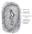
Ureter - cross-section. (WC)

Pathology of the ureter - overview
- Kidney stones.
- Ureteritis cystica.
- Urothelial carcinoma in situ - strong association with CIS in the urinary bladder.[2]
- Other urothelial neoplasias - esp. in Lynch syndrome[3] - see urothelium.
- Malakoplakia.
- Others.
Specific conditions
Ureteritis cystica
General
- Similar cystitis cystica.
- Uncommon.
- Painful.[4]
- Related to von Brunn's nests.[5]
Gross
- Smooth/round projections into the lumen.
Images:
Microscopic
Features:
- Nests of urothelium within the lamina propria with cyst formation, i.e. lumens are present.
Image:
Urothelial carcinoma
General
- Should prompt consideration of Lynch syndrome.
Microscopic
See:
Sign out
DISTAL LEFT URETER (TIE ON PROXIMAL END), EXCISION:
- HIGH-GRADE PAPILLARY UROTHELIAL CARCINOMA, NON-INVASIVE.
-- PLEASE SEE TUMOUR SUMMARY AND COMMENT.
See also
References
- ↑ URL: http://www.histology.leeds.ac.uk/urinary/ureter.php. Accessed: 31 October 2013.
- ↑ Zhou, H.; Ro, JY.; Truong, LD.; Ayala, AG.; Shen, SS. (2014). "Intraoperative frozen section evaluation of ureteral and urethral margins: studies of 203 consecutive radical cystoprostatectomy for men with bladder urothelial carcinoma.". Am J Clin Exp Urol 2 (2): 156-60. PMID 25374918.
- ↑ Crockett, DG.; Wagner, DG.; Holmäng, S.; Johansson, SL.; Lynch, HT. (May 2011). "Upper urinary tract carcinoma in Lynch syndrome cases.". J Urol 185 (5): 1627-30. doi:10.1016/j.juro.2010.12.102. PMID 21419447.
- ↑ Padilla-Fernández, B.; Díaz-Alférez, F.; Herrero-Polo, M.; Martín-Izquierdo, M.; Silva-Abuín, J.; Lorenzo-Gómez, M. (2012). "Ureteritis cystica: important consideration in the differential diagnosis of acute renal colic.". Clin Med Insights Case Rep 5: 29-33. doi:10.4137/CCRep.S9189. PMID 22474406.
- ↑ 5.0 5.1 5.2 Rothschild, JG.; Wu, G. (2011). "Ureteritis cystica: a radiologic pathologic correlation.". J Clin Imaging Sci 1: 23. doi:10.4103/2156-7514.80375. PMC 3177432. PMID 21966620. https://www.ncbi.nlm.nih.gov/pmc/articles/PMC3177432/.