Difference between revisions of "Trichoblastoma"
(create redirect) |
(→IHC) |
||
| (6 intermediate revisions by the same user not shown) | |||
| Line 1: | Line 1: | ||
# | {{ Infobox diagnosis | ||
| Name = {{PAGENAME}} | |||
| Image = Trichoepithelioma - 2 - high mag.jpg | |||
| Width = | |||
| Caption = Trichoepithelioma. [[H&E stain]]. | |||
| Micro = typically well-circumscribed cell nest in the superficial dermis, surrounding by a fibrous stroma, basaloid cells - usu. with [[peripheral palisading]] +/-surround keratin-filled cysts, fibroblasts-like cell aggregate, similar to a follicular papillae (papillary-mesenchymal body) | |||
| Subtypes = desmoplastic | |||
| LMDDx = [[basal cell carcinoma]], [[dermal cylindroma]], [[trichofolliculoma]], [[sebaceous carcinoma]], [[microcystic adnexal carcinoma]] | |||
| Stains = | |||
| IHC = | |||
| EM = | |||
| Molecular = | |||
| IF = | |||
| Gross = | |||
| Grossing = | |||
| Site = [[skin]] | |||
| Assdx = | |||
| Syndromes = | |||
| Clinicalhx = | |||
| Signs = | |||
| Symptoms = | |||
| Prevalence = uncommon | |||
| Bloodwork = | |||
| Rads = | |||
| Endoscopy = | |||
| Prognosis = benign | |||
| Other = | |||
| ClinDDx = | |||
}} | |||
'''Trichoblastoma''', also known as '''trichoepithelioma''', is an uncommon [[dermatologic neoplasms|skin tumour]]. | |||
''Trichoepithelioma'' is considered a superficial version of trichoblastoma; the World Health Organization lumps the two entities together.<ref name=Ref_Derm383>{{Ref Derm|383}}</ref> | |||
==General== | |||
*Benign. | |||
**Maligant counterpart of trichoepithelioma: [[trichilemmal carcinoma]]. | |||
*May be familial: | |||
**Multiple familial trichoepithelioma.<ref name=pmid15289313>{{Cite journal | last1 = Salhi | first1 = A. | last2 = Bornholdt | first2 = D. | last3 = Oeffner | first3 = F. | last4 = Malik | first4 = S. | last5 = Heid | first5 = E. | last6 = Happle | first6 = R. | last7 = Grzeschik | first7 = KH. | title = Multiple familial trichoepithelioma caused by mutations in the cylindromatosis tumor suppressor gene. | journal = Cancer Res | volume = 64 | issue = 15 | pages = 5113-7 | month = Aug | year = 2004 | doi = 10.1158/0008-5472.CAN-04-0307 | PMID = 15289313 }}</ref> | |||
**Brooke-Spiegler syndrome. | |||
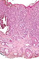
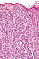
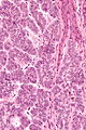
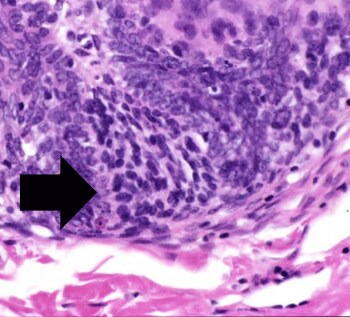
==Microscopic== | |||
Features:<ref>URL: [http://emedicine.medscape.com/article/1060049-workup#a0723 http://emedicine.medscape.com/article/1060049-workup#a0723]. Accessed on: 31 August 2011.</ref> | |||
*Well-circumscribed cell nest in the superficial dermis. | |||
*Surrounding by a fibrous stroma. | |||
*Basaloid cells with [[peripheral palisading]]. | |||
*+/-Surround keratin-filled cysts. | |||
*Fibroblasts-like cell aggregate, similar to a follicular papillae (papillary-mesenchymal body). | |||
Notes: | |||
*Very rarely an artefactual cleft - as in [[basal cell carcinoma]]. | |||
Variants: | |||
*Desmoplastic trichoblastoma. | |||
DDx: | |||
*[[Basal cell carcinoma]] - usu. mitoses, [[myxoid stroma]] and '''no''' papillary-mesenchymal bodies. | |||
*[[Dermal cylindroma]] - has hyaline stroma. | |||
*[[Trichofolliculoma]]. | |||
*[[Sebaceous carcinoma]], well-differentiated - has some cells with clear vacuolated cytoplasm. | |||
*[[Microcystic adnexal carcinoma]] - esp. desmoplastic trichoblastoma. | |||
===Images=== | |||
<gallery> | |||
Image:Trichoepithelioma - low mag.jpg | Trichoepithelioma - low mag. (WC/Nephron) | |||
Image:Trichoepithelioma - intermed mag.jpg | Trichoepithelioma - intermed. mag. (WC/Nephron) | |||
Image:Trichoepithelioma - high mag.jpg | Trichoepithelioma - high mag. (WC/Nephron) | |||
Image:Trichoepithelioma - very high mag.jpg | Trichoepithelioma - very high mag. (WC/Nephron) | |||
</gallery> | |||
<gallery> | |||
Image: Trichoepithelioma - 2 - intermed mag.jpg | Trichoepithelioma - intermed. mag. (WC) | |||
Image: Trichoepithelioma - 2 - high mag.jpg | Trichoepithelioma - high mag. (WC) | |||
Image: Trichoepithelioma - 2 - very high mag.jpg | Trichoepithelioma - very high mag. (WC) | |||
</gallery> | |||
www: | |||
*[http://img.medscape.com/pi/emed/ckb/dermatology/1048885-1055824-1060049-1348744.jpg Papillary-mesenchymal body (medscape.com)].<ref>URL: [http://emedicine.medscape.com/article/1060049-workup#a0723 http://emedicine.medscape.com/article/1060049-workup#a0723 Papillary-mesenchymal body (emedicine.medscape.com)]. Accessed on: 22 August 2012.</ref> | |||
*[http://skinpathologyatlas.com/tumors/hair/images/trichofollic-20x-pal.jpg Papillary-mesenchymal body (skinpathologyatlas.com)].<ref>URL: [http://skinpathologyatlas.com/tumors/hair/trichofollic.htm http://skinpathologyatlas.com/tumors/hair/trichofollic.htm]. Accessed on: 22 August 2012.</ref> | |||
*[http://www.dermnetnz.org/common/image.php?path=/pathology/img/t/trichoepitheliomafigure3.jpg Trichoepithelioma (dermnetnz.org)]. | |||
==IHC== | |||
Features:<ref>{{cite journal |author=Córdoba A, Guerrero D, Larrinaga B, Iglesias ME, Arrechea MA, Yanguas JI |title=Bcl-2 and CD10 expression in the differential diagnosis of trichoblastoma, basal cell carcinoma, and basal cell carcinoma with follicular differentiation |journal=Int. J. Dermatol. |volume=48 |issue=7 |pages=713–7 |year=2009 |month=July |pmid=19570076 |doi=10.1111/j.1365-4632.2009.04076.x |url=}}</ref> | |||
*CD10 +ve -- peritumoural. | |||
**Also reported in epithelium.<ref name=pmid15618927>{{cite journal |author=Yada K, Kashima K, Daa T, Kitano S, Fujiwara S, Yokoyama S |title=Expression of CD10 in basal cell carcinoma |journal=Am J Dermatopathol |volume=26 |issue=6 |pages=463–71 |year=2004 |month=December |pmid=15618927 |doi= |url=}}</ref> | |||
**Epithelial staining in BCC. | |||
*BCL2. (???) | |||
==Sign out== | |||
<pre> | |||
SKIN LESION, NOSE, BIOPSY: | |||
- TRICHOBLASTOMA, COMPLETELY EXCISED. | |||
</pre> | |||
===Micro=== | |||
The sections show well-circumscribed dermal nests of basaloid cells with peripheral palisading surrounded by a dense fibrous stroma. There is no artefactual clefting between the stroma and basaloid cell nests. Mitotic activity is minimal. Smaller hyperchromatic spindled-to-epithelioid cells in clusters (papillary-mesenchymal bodies) are found within the basaloid cells nests. | |||
The epidermis show maturation to the surface and does not have basal atypia. | |||
The lesion is completely excised in the plane of section. | |||
==See also== | |||
*[[Dermatologic neoplasms]]. | |||
==References== | |||
{{Reflist|2}} | |||
[[Category:Dermatologic neoplasms]] | |||
[[Category:Diagnosis]] | |||
Latest revision as of 17:14, 21 May 2014
| Trichoblastoma | |
|---|---|
| Diagnosis in short | |
 Trichoepithelioma. H&E stain. | |
|
| |
| LM | typically well-circumscribed cell nest in the superficial dermis, surrounding by a fibrous stroma, basaloid cells - usu. with peripheral palisading +/-surround keratin-filled cysts, fibroblasts-like cell aggregate, similar to a follicular papillae (papillary-mesenchymal body) |
| Subtypes | desmoplastic |
| LM DDx | basal cell carcinoma, dermal cylindroma, trichofolliculoma, sebaceous carcinoma, microcystic adnexal carcinoma |
| Site | skin |
|
| |
| Prevalence | uncommon |
| Prognosis | benign |
Trichoblastoma, also known as trichoepithelioma, is an uncommon skin tumour.
Trichoepithelioma is considered a superficial version of trichoblastoma; the World Health Organization lumps the two entities together.[1]
General
- Benign.
- Maligant counterpart of trichoepithelioma: trichilemmal carcinoma.
- May be familial:
- Multiple familial trichoepithelioma.[2]
- Brooke-Spiegler syndrome.
Microscopic
Features:[3]
- Well-circumscribed cell nest in the superficial dermis.
- Surrounding by a fibrous stroma.
- Basaloid cells with peripheral palisading.
- +/-Surround keratin-filled cysts.
- Fibroblasts-like cell aggregate, similar to a follicular papillae (papillary-mesenchymal body).
Notes:
- Very rarely an artefactual cleft - as in basal cell carcinoma.
Variants:
- Desmoplastic trichoblastoma.
DDx:
- Basal cell carcinoma - usu. mitoses, myxoid stroma and no papillary-mesenchymal bodies.
- Dermal cylindroma - has hyaline stroma.
- Trichofolliculoma.
- Sebaceous carcinoma, well-differentiated - has some cells with clear vacuolated cytoplasm.
- Microcystic adnexal carcinoma - esp. desmoplastic trichoblastoma.
Images
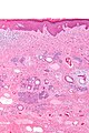
Trichoepithelioma - low mag. (WC/Nephron)
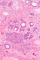
Trichoepithelioma - intermed. mag. (WC/Nephron)
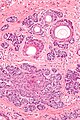
Trichoepithelioma - high mag. (WC/Nephron)
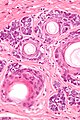
Trichoepithelioma - very high mag. (WC/Nephron)


Trichoepithelioma - intermed. mag. (WC)
Trichoepithelioma - high mag. (WC)
Trichoepithelioma - very high mag. (WC)


www:
- Papillary-mesenchymal body (medscape.com).[4]
- Papillary-mesenchymal body (skinpathologyatlas.com).[5]
- Trichoepithelioma (dermnetnz.org).
IHC
Features:[6]
- CD10 +ve -- peritumoural.
- Also reported in epithelium.[7]
- Epithelial staining in BCC.
- BCL2. (???)
Sign out
SKIN LESION, NOSE, BIOPSY:
- TRICHOBLASTOMA, COMPLETELY EXCISED.
Micro
The sections show well-circumscribed dermal nests of basaloid cells with peripheral palisading surrounded by a dense fibrous stroma. There is no artefactual clefting between the stroma and basaloid cell nests. Mitotic activity is minimal. Smaller hyperchromatic spindled-to-epithelioid cells in clusters (papillary-mesenchymal bodies) are found within the basaloid cells nests.
The epidermis show maturation to the surface and does not have basal atypia.
The lesion is completely excised in the plane of section.
See also
References
- ↑ Busam, Klaus J. (2009). Dermatopathology: A Volume in the Foundations in Diagnostic Pathology Series (1st ed.). Saunders. pp. 383. ISBN 978-0443066542.
- ↑ Salhi, A.; Bornholdt, D.; Oeffner, F.; Malik, S.; Heid, E.; Happle, R.; Grzeschik, KH. (Aug 2004). "Multiple familial trichoepithelioma caused by mutations in the cylindromatosis tumor suppressor gene.". Cancer Res 64 (15): 5113-7. doi:10.1158/0008-5472.CAN-04-0307. PMID 15289313.
- ↑ URL: http://emedicine.medscape.com/article/1060049-workup#a0723. Accessed on: 31 August 2011.
- ↑ URL: http://emedicine.medscape.com/article/1060049-workup#a0723 Papillary-mesenchymal body (emedicine.medscape.com). Accessed on: 22 August 2012.
- ↑ URL: http://skinpathologyatlas.com/tumors/hair/trichofollic.htm. Accessed on: 22 August 2012.
- ↑ Córdoba A, Guerrero D, Larrinaga B, Iglesias ME, Arrechea MA, Yanguas JI (July 2009). "Bcl-2 and CD10 expression in the differential diagnosis of trichoblastoma, basal cell carcinoma, and basal cell carcinoma with follicular differentiation". Int. J. Dermatol. 48 (7): 713–7. doi:10.1111/j.1365-4632.2009.04076.x. PMID 19570076.
- ↑ Yada K, Kashima K, Daa T, Kitano S, Fujiwara S, Yokoyama S (December 2004). "Expression of CD10 in basal cell carcinoma". Am J Dermatopathol 26 (6): 463–71. PMID 15618927.