Difference between revisions of "Thymoma"
Jump to navigation
Jump to search
| Line 1: | Line 1: | ||
{{ Infobox diagnosis | {{ Infobox diagnosis | ||
| Name = {{PAGENAME}} | | Name = {{PAGENAME}} | ||
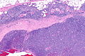
| Image = | | Image = Thymoma type B1 -- intermed mag.jpg | ||
| Width = | | Width = | ||
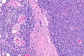
| Caption = | | Caption = Thymoma (right of image) and remnant of normal thymus (left of image). [[H&E stain]]. (WC) | ||
| Synonyms = | | Synonyms = | ||
| Micro = | | Micro = | ||
| Line 75: | Line 75: | ||
Note: | Note: | ||
*Neoplastic cells derived from the thymus with cytologic features of malignancy are [[thymic carcinoma]]s. | *Neoplastic cells derived from the thymus with cytologic features of malignancy are [[thymic carcinoma]]s. | ||
==Gross== | ==Gross== | ||
| Line 104: | Line 97: | ||
===Images=== | ===Images=== | ||
<gallery> | <gallery> | ||
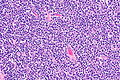
Image:Thymoma_B1_ | Image:Thymoma_type_B1_(1).JPG | Thymoma Type B1. (WC/KGH) | ||
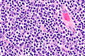
Image:Thymoma_B1_(2).JPG | Thymoma Type B1. (WC/KGH) | |||
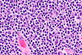
Image:Thymoma_B1_(3)_CK_CAM5-2.JPG | Thymoma Type B1 - CAM5.2. (WC/KGH) | |||
</gallery> | |||
<gallery> | |||
Image: Thymoma type B1 -- low mag.jpg | Thymoma B1 - low mag. | |||
Image: Thymoma type B1 -- intermed mag.jpg | Thymoma B1 - intermed. mag. | |||
Image: Thymoma type B1 -- high mag.jpg | Thymoma B1 - high mag. | |||
Image: Thymoma type B1 -- very high mag.jpg | Thymoma B1 - very high mag. | |||
Image: Thymoma type B1 - alt -- very high mag.jpg | Thymoma B1 - very high mag. | |||
</gallery> | </gallery> | ||
===Staging=== | ===Staging=== | ||
Revision as of 02:32, 21 December 2015
| Thymoma | |
|---|---|
| Diagnosis in short | |
 Thymoma (right of image) and remnant of normal thymus (left of image). H&E stain. (WC) | |
| Subtypes | A, AB, B1, B2, B3 |
| LM DDx | lymphoma, squamous cell carcinoma (esp. squamous cell carcinoma of the lung, head and neck squamous cell carcinoma), follicular dendritic cell sarcoma |
| Staging | thymus staging |
| Site | thymus |
|
| |
| Associated Dx | myasthenia gravis |
| Prevalence | uncommon overall, common for thymic tumours |
| Prognosis | benign |
| Clin. DDx | mediastinal mass |
| Treatment | excision |
Thymoma is a common tumour of the thymus.
General
- Strong association with autoimmune disease, esp. myasthenia gravis.
Classification
The WHO published a widely used system - WHO classification:[1]
Type A
- AKA Spindle cell or medullary.
- Arise from medullary epithelial cells.
- Good prognosis.
IHC:
- Usu. keratin+.
Type AB
- Like Type A... but with foci of lymphocytes.
Type B1
- Near normal, expanded cortex.
Lesion consists of:
- >2/3 lymphocytes, <1/3 cortical epithelial cells.
Type B2
- Neoplastic cells with some resemblance to cortical epithelial cells.
- Epithelioid cells with distinct nucleoli.
- May be perivascular.
- Large population of lymphocytes.
Lesion consists of:
- <2/3 but >1/3 lymphocytes, >1/3 but <2/3 cortical epithelial cells.
Notes:
- Most common B type.
Type B3
- Neoplastic cells with some resemblance to cortical epithelial cells.
- Polygonal/round shape.
- Form sheets (of cells) - key feature.
- Lymphocytes - less than in Type B2.
- AKA well-differentiated thymic carcinoma.
Lesion consists of:
- <1/3 lymphocytes, >2/3 cortical epithelial cells.
Note:
- Neoplastic cells derived from the thymus with cytologic features of malignancy are thymic carcinomas.
Gross
- Light brown/tan.
- Encapsulated.
Image:
Microscopic
Features:
- Lymphocytes.
- Epithelial cells.
- Spindle cells - Type A.
- Epithelioid cells - Type B.
DDx:
Images
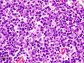
Thymoma Type B1. (WC/KGH)
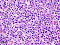
Thymoma Type B1. (WC/KGH)
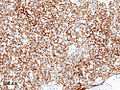
Thymoma Type B1 - CAM5.2. (WC/KGH)
.JPG)
.JPG)
_CK_CAM5-2.JPG)
Thymoma B1 - low mag.
Thymoma B1 - intermed. mag.
Thymoma B1 - high mag.
Thymoma B1 - very high mag.
Thymoma B1 - very high mag.


Staging
Main article: Thymic staging
IHC
A panel:
- TdT, CD1a, CD3, CD5, CD20, Ki-67, CD117, p63, CK5/6.
Sign out
A. Lymph Node, Station 6, Lymphadenectomy:
- One benign lymph node (0/1).

B. Submitted as "Anterior Mediastinal Tumour (Thymus)", Excision:
- Thymoma, WHO type B2.
- Modified Masaoka stage IIa.
- Three benign lymph nodes (0/3).
- Rim of benign thymus.
- Please see synoptic report.
See also
References
- ↑ Mills, Stacey E; Carter, Darryl; Greenson, Joel K; Oberman, Harold A; Reuter, Victor E (2004). Sternberg's Diagnostic Surgical Pathology (4th ed.). Lippincott Williams & Wilkins. pp. 1264. ISBN 978-0781740517.
- ↑ Adam P, Hakroush S, Hofmann I, Reidenbach S, Marx A, Ströbel P (June 2014). "Thymoma with loss of keratin expression (and giant cells): a potential diagnostic pitfall". Virchows Arch.. doi:10.1007/s00428-014-1606-6. PMID 24923897.
- ↑ Viti, A.; Bertolaccini, L.; Cavallo, A.; Fortunato, M.; Bianchi, A.; Terzi, A. (Sep 2014). "18-Fluorine fluorodeoxyglucose positron emission tomography in the pretreatment evaluation of thymic epithelial neoplasms: a metabolic biopsy confirmed by Ki-67 expression.". Eur J Cardiothorac Surg 46 (3): 369-74; discussion 374. doi:10.1093/ejcts/ezu030. PMID 24585679.