Difference between revisions of "TFE3-rearranged renal cell carcinoma"
Jump to navigation
Jump to search
(redirect) |
(+infobox) |
||
| Line 1: | Line 1: | ||
{{ Infobox diagnosis | |||
| Name = {{PAGENAME}} | |||
| Image = Xp11.2_translocation_renal_cell_carcinoma_-_high_mag.jpg | |||
| Width = | |||
| Caption = Xp11.2 translocation carcinoma. [[H&E stain]]. | |||
| Micro = large cells with clear or eosinophilic cytoplasm, calcification (classic histomorphologic feature), +/-papillae, +/-nests, +/-[[psammoma bodies]] (common), +/-[[hyaline bodies]] (common) | |||
| Subtypes = | |||
| LMDDx = [[clear cell renal cell carcinoma]], [[papillary renal cell carcinoma]], [[epithelioid angiomyolipoma]], [[clear cell papillary renal cell carcinoma]] | |||
| Stains = | |||
| IHC = TFE3 +ve (nucleus), CD10 +ve, vimentin +ve, CK7 -ve (usu.) | |||
| EM = | |||
| Molecular = [[translocation]] involving TFE3, e.g. t(X;1)(p11.2;q21) | |||
| IF = | |||
| Gross = | |||
| Grossing = | |||
| Site = [[kidney]] - see [[kidney tumours]] | |||
| Assdx = | |||
| Syndromes = | |||
| Clinicalhx = | |||
| Signs = | |||
| Symptoms = | |||
| Prevalence = rare | |||
| Bloodwork = | |||
| Rads = | |||
| Endoscopy = | |||
| Prognosis = poor | |||
| Other = | |||
| ClinDDx = other [[kidney tumours]] | |||
}} | |||
'''Renal tumour with Xp11.2 translocation''', also '''Xp11.2 translocation carcinoma''', is an uncommon [[malignant]] [[kidney tumour]]. | '''Renal tumour with Xp11.2 translocation''', also '''Xp11.2 translocation carcinoma''', is an uncommon [[malignant]] [[kidney tumour]]. | ||
Revision as of 16:24, 23 November 2013
| TFE3-rearranged renal cell carcinoma | |
|---|---|
| Diagnosis in short | |
|
Xp11.2 translocation carcinoma. H&E stain. | |
|
| |
| LM | large cells with clear or eosinophilic cytoplasm, calcification (classic histomorphologic feature), +/-papillae, +/-nests, +/-psammoma bodies (common), +/-hyaline bodies (common) |
| LM DDx | clear cell renal cell carcinoma, papillary renal cell carcinoma, epithelioid angiomyolipoma, clear cell papillary renal cell carcinoma |
| IHC | TFE3 +ve (nucleus), CD10 +ve, vimentin +ve, CK7 -ve (usu.) |
| Molecular | translocation involving TFE3, e.g. t(X;1)(p11.2;q21) |
| Site | kidney - see kidney tumours |
|
| |
| Prevalence | rare |
| Prognosis | poor |
| Clin. DDx | other kidney tumours |
Renal tumour with Xp11.2 translocation, also Xp11.2 translocation carcinoma, is an uncommon malignant kidney tumour.
General
- Defined by the presence of a fusion gene formed with TFE3 @ Xp11.2.
- TFE3 is the gene involved in the translocation seen in alveolar soft part sarcoma (ASPS).
- Poor prognosis ~ 50% present at stage IV, majority of lymph node metastases.
- ~1/3 of childhood RCC.[1]
Microscopic
Features:[2]
- Large cells.
- Clear or eosinophilic cytoplasm.
- Papillae or nests.
- Psammoma bodies - common.[3]
- Calcification is considered the classic histomorphologic feature.
- Hyaline bodies - common.
Notes:
- Looks clear cell RCC or papillary RCC or a hybrid between the two.
- May resemble alveolar soft part sarcoma.
DDx:
- Clear cell RCC.
- Papillary RCC.
- Epithelioid angiomyolipoma.
- Clear cell papillary renal cell carcinoma.
Images
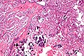
Xp11.2 translocation RCC - intermed. mag. (WC/Nephron)
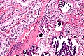
Xp11.2 translocation RCC - high mag. (WC/Nephron)
www:
IHC
Others:
- HMB-45 & Melan A -ve.
- Positive in epithelioid angiomyolipoma.
Molecular
- Translocation involving TFE3, e.g. t(X;1)(p11.2;q21).[2]
See also
References
- ↑ Argani, P.; Olgac, S.; Tickoo, SK.; Goldfischer, M.; Moch, H.; Chan, DY.; Eble, JN.; Bonsib, SM. et al. (Aug 2007). "Xp11 translocation renal cell carcinoma in adults: expanded clinical, pathologic, and genetic spectrum.". Am J Surg Pathol 31 (8): 1149-60. doi:10.1097/PAS.0b013e318031ffff. PMID 17667536.
- ↑ 2.0 2.1 2.2 Humphrey, Peter A; Dehner, Louis P; Pfeifer, John D (2008). The Washington Manual of Surgical Pathology (1st ed.). Lippincott Williams & Wilkins. pp. 285. ISBN 978-0781765275.
- ↑ Prasad, SR.; Humphrey, PA.; Catena, JR.; Narra, VR.; Srigley, JR.; Cortez, AD.; Dalrymple, NC.; Chintapalli, KN.. "Common and uncommon histologic subtypes of renal cell carcinoma: imaging spectrum with pathologic correlation.". Radiographics 26 (6): 1795-806; discussion 1806-10. doi:10.1148/rg.266065010. PMID 17102051.
- ↑ He, H.; Zhou, GX.; Zhou, M.; Chen, L. (Sep 2011). "The distinction of clear cell carcinoma of the female genital tract, clear cell renal cell carcinoma, and translocation-associated renal cell carcinoma: an immunohistochemical study using tissue microarray.". Int J Gynecol Pathol 30 (5): 425-30. doi:10.1097/PGP.0b013e318214dd4f. PMID 21804394.