Steatosis
Steatosis is a fatty change in the liver associated with a number of underlying causes.
It is also known as fatty liver.
General
Classification
Can be divided into:
- Macrovesicular steatosis.
- Common.
- Microvesicular steatosis.
- Rare.
- Potentially life threatening.[1]
Note:
- It is considered technically incorrect to say the liver, in steatosis/steatohepatitis, contains adipocytes; they are lipid-laden hepatocytes,[2] despite that:
- Histologically, these cells look like adipocytes.
- Lipid-laden hepatocytes have gene activations suggestive of adipogenic-like transformation.[3]
Etiology
Microvesicular steatosis
Microvesicular steatosis DDx:[4]
- Acute fatty liver of pregnancy,
- Reye's syndrome.
- Drug toxicity:
- Sodium valproate toxicity.
- High-dose tetracycline toxicity.
- Jamaican vomiting sickness.
- Congenital defects of urea cycle enzymes.
Less common causes:
- Alcoholism.
- Hepatitis D.
- Weird stuff:
- Congenital defects of fatty acid beta oxidation,
- Cholesterol ester storage disease,
- Wolman disease and Alpers syndrome.
The classic causes of microvesicular steatosis are:[5]
- Fatty liver of pregnancy.
- Aspirin (Reye's syndrome).
- Tetracycline.
It was once thought that all other causes of fatty liver produce macrovesicular steatosis.
Macrovesicular steatosis
Can sometimes be divided into centrilobular predominant and periportal predominant.[6]
Centrilobular predominant (zone III) - DOA:[6]
- Diabetes mellitus.
- Obesity, non-alcoholic steatohepatitis (NASH).
- Alcoholic liver disease, alcoholic steatohepatitis (ASH).
Periportal predominant (zone I) - TAPES:[6]
- Total parenteral nutrition (TPN).
- AIDS.
- Phosphorus poisoning.
- Exogenous steroids.
- Starvation.[7]
Notes:
- HCV genotype 3 is reported to cause periportal steatosis.[8]
- Donor livers with more macrovescicular steatosis = worse outcome.
- More than 30% means the liver is undesirable for transplantation.[9]
Microscopic
Features - macrovesicular steatosis.
- One large vacuoles - similar to mature adipose tissue.
- Nucleus is eccentric.
Features - microvesicular steatosis.
- Multiple small (clear) cytoplasmic vacuoles - similar to brown fat, as seen in a hibernoma.
- Nucleus is central.[10]
Grading
Quantity of fat is usually given as a percentage and graded mild, moderate, or marked.
- Mild <33%, moderate >33% & <66%, marked >66%.[11]
Images
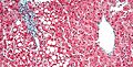
Periportal steatosis. (WC/Nephron)

Image:
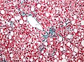
Centrilobular steatosis. (WC/Nephron)

See also
References
- ↑ Jolly, RA.; Ciurlionis, R.; Morfitt, D.; Helgren, M.; Patterson, R.; Ulrich, RG.; Waring, JF.. "Microvesicular steatosis induced by a short chain fatty acid: effects on mitochondrial function and correlation with gene expression.". Toxicol Pathol 32 Suppl 2: 19-25. PMID 15503661.
- ↑ Guindi, M. September 2009.
- ↑ URL: http://www.jci.org/articles/view/20513/version/1. Accessed on: 23 September 2009.
- ↑ Hautekeete ML, Degott C, Benhamou JP (1990). "Microvesicular steatosis of the liver". Acta Clin Belg 45 (5): 311–26. PMID 2177300.
- ↑ http://www.mailman.srv.ualberta.ca/pipermail/patho-l/1996-June/001788.html
- ↑ 6.0 6.1 6.2 Steatosis. pathconsultddx.com. URL: http://www.pathconsultddx.com/pathCon/diagnosis?pii=S1559-8675%2806%2970840-3. Accessed on: 2 Sep 2009.
- ↑ Nagy, I.; Németh, J.; Lászik, Z. (Jan 2000). "Effect of L-aminocarnitine, an inhibitor of mitochondrial fatty acid oxidation, on the exocrine pancreas and liver in fasted rats.". Pharmacol Res 41 (1): 9-17. doi:10.1006/phrs.1999.0565. PMID 10600264.
- ↑ Yoon EJ, Hu KQ. Hepatitis C virus (HCV) infection and hepatic steatosis. Int J Med Sci. 2006;3(2):53-6. Epub 2006 Apr 1. PMID 16614743. Avialable at: http://www.pubmedcentral.nih.gov/articlerender.fcgi?artid=1415843. Accessed on: September 9, 2009.
- ↑ STC. 6 December 2010.
- ↑ STC. 6 December 2010.
- ↑ Guindi, M. September 17, 2009.