Difference between revisions of "Spleen"
(+littoral cell angioma) |
|||
| (69 intermediate revisions by the same user not shown) | |||
| Line 1: | Line 1: | ||
[[Image:Gray1188.png|thumb|300px|Spleen.]] | |||
The '''spleen''' is a forgotten organ. | The '''spleen''' is a forgotten organ. | ||
== | =Normal histology= | ||
*Capsule. | |||
*Red pulp - [[red blood cells]]. | |||
*White pulp - white blood cells. | |||
*Marginal zone - between red pulp and white pulp.<ref>{{Ref WMSP|589}}</ref> | |||
*Mononucleosis (EBV infection). | ===Images=== | ||
<gallery> | |||
Image:Splenic_hamartoma_-_very_low_mag.jpg | Normal spleen (left) and splenic hamartoma (right). H&E stain. (WC) | |||
</gallery> | |||
==Sign out== | |||
===Micro=== | |||
Sections show unremarkable splenic parenchyma. The nodules of white pulp are well-spaced and have germinal center formation. The red pulp has a normal vascularity and does not appear to be expanded. No nuclear atypia is apparent. | |||
=Gross pathology= | |||
The [[grossing]] of the spleen is dealt with in [[splenectomy grossing]]. | |||
===Overview of classic gross findings=== | |||
{| class = "wikitable sortable" | |||
! Terminology | |||
! Etiology | |||
! Description | |||
! Other | |||
|- | |||
|Ruptured spleen | |||
| trauma | |||
| hemorrhagic, capsule disrupted | |||
| possible assocations [[mononucleosis]], medical procedure - esp. colonoscopy,<ref name=pmid22889306>{{Cite journal | last1 = Aubrey-Bassler | first1 = FK. | last2 = Sowers | first2 = N. | title = 613 cases of splenic rupture without risk factors or previously diagnosed disease: a systematic review. | journal = BMC Emerg Med | volume = 12 | issue = 1 | pages = 11 | month = Aug | year = 2012 | doi = 10.1186/1471-227X-12-11 | PMID = 22889306 }}</ref> others | |||
|- | |||
|"Lardaceous spleen"<ref name=Ref_Klatt91>{{Ref Klatt|91}}</ref> | |||
| [[amyloidosis]] | |||
| waxy, pale, grey | |||
| also see ''sago spleen'' | |||
|- | |||
|"Sago spleen" | |||
| [[amyloidosis]] | |||
| nodular, tapioca-like appearance | |||
| also see lardaceous spleen | |||
|- | |||
|[[Splenic infarct]] | |||
| vascular occulsion | |||
| wedge shaped -- periphery | |||
| - | |||
|- | |||
|} | |||
Images: | |||
*[http://www.pathguy.com/lectures/sagolard.gif Amyloidosis of the spleen - schematic (pathguy.com)]. | |||
*[http://commons.wikimedia.org/wiki/File:Tapioca_pudding-3.jpg Tapioca pudding (WC)]. | |||
=Splenic enlargement= | |||
:''Splenomegaly'' redirects here.'' | |||
*[[Portal hypertension]] (often due to [[cirrhosis]]). | |||
*Lymphoma - see below. | |||
*Mononucleosis ([[EBV]] infection). | |||
*[[Hemophagocytic syndrome]].<ref name=pmid20127750>{{Cite journal | last1 = Maakaroun | first1 = NR. | last2 = Moanna | first2 = A. | last3 = Jacob | first3 = JT. | last4 = Albrecht | first4 = H. | title = Viral infections associated with haemophagocytic syndrome. | journal = Rev Med Virol | volume = 20 | issue = 2 | pages = 93-105 | month = Mar | year = 2010 | doi = 10.1002/rmv.638 | PMID = 20127750 }} | *[[Hemophagocytic syndrome]].<ref name=pmid20127750>{{Cite journal | last1 = Maakaroun | first1 = NR. | last2 = Moanna | first2 = A. | last3 = Jacob | first3 = JT. | last4 = Albrecht | first4 = H. | title = Viral infections associated with haemophagocytic syndrome. | journal = Rev Med Virol | volume = 20 | issue = 2 | pages = 93-105 | month = Mar | year = 2010 | doi = 10.1002/rmv.638 | PMID = 20127750 }} | ||
</ref> | </ref> | ||
== | ==Lymphoid neoplasms of the spleen== | ||
* | Lymphomas of the spleen in order of prevalence - in a series of 115 cases:<ref name=pmid22924843>{{Cite journal | last1 = Shimizu-Kohno | first1 = K. | last2 = Kimura | first2 = Y. | last3 = Kiyasu | first3 = J. | last4 = Miyoshi | first4 = H. | last5 = Yoshida | first5 = M. | last6 = Ichikawa | first6 = R. | last7 = Niino | first7 = D. | last8 = Ohshima | first8 = K. | title = Malignant lymphoma of the spleen in Japan: a clinicopathological analysis of 115 cases. | journal = Pathol Int | volume = 62 | issue = 9 | pages = 577-82 | month = Sep | year = 2012 | doi = 10.1111/j.1440-1827.2012.02844.x | PMID = 22924843 }}</ref> | ||
*Capsule of the spleen is white - resembles sugar-coating | #[[Diffuse large B-cell lymphoma]] (DLBCL). | ||
* | #[[Splenic marginal zone lymphoma]] (SMZL).<ref name=pmid20350661>{{Cite journal | last1 = Bennett | first1 = M. | last2 = Schechter | first2 = GP. | title = Treatment of splenic marginal zone lymphoma: splenectomy versus rituximab. | journal = Semin Hematol | volume = 47 | issue = 2 | pages = 143-7 | month = Apr | year = 2010 | doi = 10.1053/j.seminhematol.2010.01.004 | PMID = 20350661 }}</ref> | ||
#[[Follicular lymphoma]]. | |||
#Splenic B-cell lymphoma, unclassifiable. | |||
#[[Peripheral T-cell lymphoma]], not otherwise specified. | |||
Less common lymphoid neoplasms of the spleen: | |||
*[[Hairy cell leukemia]]. | |||
*Splenic diffuse red pulp small B cell lymphoma.<ref name=pmid20677173>{{cite journal |author=Baseggio L, Traverse-Glehen A, Callet-Bauchu E, ''et al.'' |title=Relevance of a scoring system including CD11c expression in the identification of splenic diffuse red pulp small B-cell lymphoma (SRPL) |journal=Hematol Oncol |volume=29 |issue=1 |pages=47–51 |year=2011 |month=March |pmid=20677173 |doi=10.1002/hon.957 |url=}}</ref> | |||
==DDx by compartment== | |||
===White pulp malignant=== | |||
*[[Diffuse large B-cell lymphoma]]. | |||
*[[Splenic marginal zone lymphoma]]. | |||
*[[Follicular lymphoma]]. | |||
===Red pulp benign=== | |||
*[[Polycythemia rubra vera]] (AKA polycythemia vera). | |||
*Outflow obstruction. | |||
**[[Cirrhosis]]. | |||
**[[Budd-Chiari syndrome]] (AKA hepatic vein obstruction). | |||
**Splenic vein thrombosis. | |||
*[[Congestive heart failure]]. | |||
*[[Acute splenitis]]. | |||
===Red pulp malignant=== | |||
*[[Hairy cell leukemia]]. | |||
*Acute leukemia.<ref name=Ref_WMSP_596>{{Ref WMSP|596}}</ref> | |||
*[[Hepatosplenic T-cell lymphoma]]<ref name=Ref_WMSP595>{{Ref WMSP|595}}</ref> (e.g. Hepatosplenic γδ T-cell lymphoma). | |||
=Specific disorders= | |||
==Splenic laceration== | |||
{{Main|Splenic laceration}} | |||
==Hyaloserositis of the spleen== | |||
*[[AKA]] ''sugar-coated spleen''. | |||
===General=== | |||
*Benign. | |||
*Common [[autopsy]] finding. | |||
===Gross=== | |||
*Capsule of the spleen is white - resembles sugar-coating.<ref>URL: [http://www.drugs.com/dict/sugar-coated-spleen.html http://www.drugs.com/dict/sugar-coated-spleen.html]. Accessed on: 1 September 2010.</ref> | |||
===Microscopic=== | |||
Features: | |||
*Hyaline material adherent to splenic capsule. | |||
**Hyaline material = pink acellular crap on a [[H&E stain]]. | |||
Image( | ====Images==== | ||
<gallery> | |||
Image:Spleen_hyaloserositis_-_intermed_mag.jpg | Hyaloserositis - intermed. mag. (WC) | |||
Image:Spleen_hyaloserositis_-b-_high_mag.jpg | Hyaloserositis - high mag. (WC) | |||
</gallery> | |||
==Mononucleosis== | ==Mononucleosis== | ||
===General=== | ===General=== | ||
*[[EBV]] infection. | *[[EBV]] infection. | ||
*Massive splenic enlargement. | *+/-Massive splenic enlargement. | ||
Clinical: | |||
*Monospot test +ve. | |||
===Microscopic=== | ===Microscopic=== | ||
Features: | Features: | ||
*Atypical lymphoid cells. | *Atypical lymphoid cells. | ||
**Abundant basophilic cytoplasm. | |||
**Cells indented by adjacent RBCs on blood smears.<ref name=utah_endo>URL: [http://library.med.utah.edu/WebPath/EXAM/IMGQUIZ/hpfrm.html http://library.med.utah.edu/WebPath/EXAM/IMGQUIZ/hpfrm.html]. Accessed on: 4 December 2011.</ref> | |||
Images: | |||
*[http://library.med.utah.edu/WebPath/jpeg5/HEME013.jpg Mononucleosis (med.utah.edu)].<ref name=utah_endo>URL: [http://library.med.utah.edu/WebPath/EXAM/IMGQUIZ/hpfrm.html http://library.med.utah.edu/WebPath/EXAM/IMGQUIZ/hpfrm.html]. Accessed on: 4 December 2011.</ref> | |||
*[http://path.upmc.edu/cases/case37.html Mononucleosis - several images (upmc.edu)]. | |||
===Flow cytometry=== | |||
*CD8 >>> CD4.<ref>URL: [http://path.upmc.edu/cases/case37/gross.html http://path.upmc.edu/cases/case37/gross.html]. Accessed on: 2 January 2012.</ref> | |||
==Littoral cell angioma== | |||
:For ''angioma'' see ''[[vascular malformations]]''. | |||
{{Main|Littoral cell angioma}} | |||
==Splenic hamartoma== | |||
{{Main|Splenic hamartoma}} | |||
==Splenic infarct== | |||
*[[AKA]] ''splenic [[infarct]]ion''. | |||
*[[AKA]] ''infarction of the spleen''. | |||
===General=== | |||
Classic textbook causes:<ref name=pmid20928991>{{Cite journal | last1 = Lawrence | first1 = YR. | last2 = Pokroy | first2 = R. | last3 = Berlowitz | first3 = D. | last4 = Aharoni | first4 = D. | last5 = Hain | first5 = D. | last6 = Breuer | first6 = GS. | last7 = Osler | first7 = W. | title = Splenic infarction: an update on William Osler's observations. | journal = Isr Med Assoc J | volume = 12 | issue = 6 | pages = 362-5 | month = Jun | year = 2010 | doi = | PMID = 20928991 | URL = http://www.ima.org.il/imaj/dynamic/web/ArtFromPubmed.asp?year=2010&month=06&page=362 }}</ref> | |||
*Septic embolus due to [[bacterial endocarditis]]. | |||
*[[Sickle cell disease]]. | |||
Usual causes:<ref name=pmid20928991/> | |||
*Hematologic malignancy. | |||
*Intracardiac thrombus. | |||
*Bacterial endocarditis. | |||
Clinical:<ref name=pmid20928991/> | |||
*Left upper quadrant pain ~ 1/3 of cases. | |||
*Fever ~ 1/3 of cases. | |||
*Leukocytosis ~ 1/2 of cases. | |||
===Gross=== | |||
*Classically wedge-shaped; triangular on section. | |||
**The base of the triangle runs along the surface. | |||
**The apex points to the obstructed vessel that lead to the infarct. | |||
===Microscopic=== | |||
:''See [[necrosis]]''. | |||
=Weird stuff= | |||
*Dendritic cell tumours.<ref name=Ref_WMSP_596>{{Ref WMSP|596}}</ref> | |||
**Interdigitating dendritic cell tumour. | |||
**Follicular dendritic cell tumour. | |||
==Follicular dendritic cell tumour== | |||
*[[AKA]] ''follicular dendritic cell sarcoma''. | |||
{{Main|Follicular dendritic cell sarcoma}} | |||
== | ==Hepatosplenic T-cell lymphoma== | ||
*Abbreviated ''HSTL''. | |||
===General=== | ===General=== | ||
*Rare. | *Rare. | ||
* | *Prognosis: poor. | ||
Subtypes:<ref name=webpath_HSTL/> | |||
#''Hepatosplenic γδ T-cell lymphoma''. | |||
#*May be written ''Hepatosplenic gamma/delta T-cell lymphoma''. | |||
#''Hepatosplenic αβ T-cell lymphoma''. | |||
#*May be written ''Hepatosplenic alpha/beta T-cell lymphoma''. | |||
Clinical triad: | |||
#Hepatosplenomegaly. | |||
#Cytopenias ([[anemia]], thrombocytopenia). | |||
#Sinusoidal tropism. | |||
===Microscopic=== | ===Microscopic=== | ||
Features:<ref name= | Features:<ref name=Ref_WMSP595>{{Ref WMSP|595}}</ref><ref name=webpath_HSTL>URL: [http://www.webpathology.com/image.asp?case=378&n=15 http://www.webpathology.com/image.asp?case=378&n=15]. Accessed on: 22 December 2011.</ref> | ||
* | *Small cell lymphoma/Intermediate cell lymphoma. | ||
**+/-"Folded" nuclei. | |||
DDx: | |||
*[[Hairy cell leukemia]]. | |||
Images: | |||
*[http://www.webpathology.com/image.asp?n=14&Case=378 HSTL - low mag. (webpathology.com)]. | |||
*[http://www.webpathology.com/image.asp?case=378&n=15 HSTL - high mag. (webpathology.com)]. | |||
===IHC=== | |||
Features:<ref name=Ref_WMSP595>{{Ref WMSP|595}}</ref> | |||
*CD4 -ve. | |||
*CD8 -ve. | |||
*NK cell-associated antigens +ve:<ref name=pmid9191001/> | |||
**[[CD56]],<ref name=pmid15458516>{{Cite journal | last1 = Niitsu | first1 = N. | last2 = Kohri | first2 = M. | last3 = Togano | first3 = T. | last4 = Nakamine | first4 = H. | last5 = Nakamura | first5 = S. | last6 = Iwabuchi | first6 = K. | last7 = Higashihara | first7 = M. | title = Development of hepatosplenic gammadelta T-cell lymphoma with pancytopenia during early pregnancy: a case report and review of the literature. | journal = Eur J Haematol | volume = 73 | issue = 5 | pages = 367-71 | month = Nov | year = 2004 | doi = 10.1111/j.1600-0609.2004.00300.x | PMID = 15458516 }}</ref> CD11c, CD16. | |||
*Effector proteins +ve.<ref name=pmid9191001>{{Cite journal | last1 = Salhany | first1 = KE. | last2 = Feldman | first2 = M. | last3 = Kahn | first3 = MJ. | last4 = Peritt | first4 = D. | last5 = Schretzenmair | first5 = RD. | last6 = Wilson | first6 = DM. | last7 = DiPaola | first7 = RS. | last8 = Glick | first8 = AD. | last9 = Kant | first9 = JA. | title = Hepatosplenic gammadelta T-cell lymphoma: ultrastructural, immunophenotypic, and functional evidence for cytotoxic T lymphocyte differentiation. | journal = Hum Pathol | volume = 28 | issue = 6 | pages = 674-85 | month = Jun | year = 1997 | doi = | PMID = 9191001 }}</ref> | |||
**Perforin, granzyme B, TIA-1, Fas ligand. | |||
==Myelolipoma of the spleen== | |||
{{Main|Myelolipoma}} | |||
==Immune thrombocytopenic purpura== | |||
{{Main|Immune thrombocytopenic purpura}} | |||
Spleens are removed for this. | |||
==Splenogonadal fusion== | |||
{{Main|Splenogonadal fusion}} | |||
=See also= | |||
*[[Haematopathology]]. | *[[Haematopathology]]. | ||
*[[Liver pathology]]. | *[[Liver pathology]]. | ||
=References= | |||
{{reflist|2}} | {{reflist|2}} | ||
=External links= | |||
*[http://www.rcsed.ac.uk/journal/vol47_1/47100010.html Atraumatic rupture of the spleen in adults (rcsed.ac.uk)]. | *[http://www.rcsed.ac.uk/journal/vol47_1/47100010.html Atraumatic rupture of the spleen in adults (rcsed.ac.uk)]. | ||
[[Category:Haematopathology]] | [[Category:Haematopathology]] | ||
[[Category:Spleen]] | |||
Latest revision as of 12:50, 7 September 2016

The spleen is a forgotten organ.
Normal histology
- Capsule.
- Red pulp - red blood cells.
- White pulp - white blood cells.
- Marginal zone - between red pulp and white pulp.[1]
Images
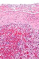
Normal spleen (left) and splenic hamartoma (right). H&E stain. (WC)

Sign out
Micro
Sections show unremarkable splenic parenchyma. The nodules of white pulp are well-spaced and have germinal center formation. The red pulp has a normal vascularity and does not appear to be expanded. No nuclear atypia is apparent.
Gross pathology
The grossing of the spleen is dealt with in splenectomy grossing.
Overview of classic gross findings
| Terminology | Etiology | Description | Other |
|---|---|---|---|
| Ruptured spleen | trauma | hemorrhagic, capsule disrupted | possible assocations mononucleosis, medical procedure - esp. colonoscopy,[2] others |
| "Lardaceous spleen"[3] | amyloidosis | waxy, pale, grey | also see sago spleen |
| "Sago spleen" | amyloidosis | nodular, tapioca-like appearance | also see lardaceous spleen |
| Splenic infarct | vascular occulsion | wedge shaped -- periphery | - |
Images:
Splenic enlargement
- Splenomegaly redirects here.
- Portal hypertension (often due to cirrhosis).
- Lymphoma - see below.
- Mononucleosis (EBV infection).
- Hemophagocytic syndrome.[4]
Lymphoid neoplasms of the spleen
Lymphomas of the spleen in order of prevalence - in a series of 115 cases:[5]
- Diffuse large B-cell lymphoma (DLBCL).
- Splenic marginal zone lymphoma (SMZL).[6]
- Follicular lymphoma.
- Splenic B-cell lymphoma, unclassifiable.
- Peripheral T-cell lymphoma, not otherwise specified.
Less common lymphoid neoplasms of the spleen:
- Hairy cell leukemia.
- Splenic diffuse red pulp small B cell lymphoma.[7]
DDx by compartment
White pulp malignant
Red pulp benign
- Polycythemia rubra vera (AKA polycythemia vera).
- Outflow obstruction.
- Cirrhosis.
- Budd-Chiari syndrome (AKA hepatic vein obstruction).
- Splenic vein thrombosis.
- Congestive heart failure.
- Acute splenitis.
Red pulp malignant
- Hairy cell leukemia.
- Acute leukemia.[8]
- Hepatosplenic T-cell lymphoma[9] (e.g. Hepatosplenic γδ T-cell lymphoma).
Specific disorders
Splenic laceration
Hyaloserositis of the spleen
- AKA sugar-coated spleen.
General
- Benign.
- Common autopsy finding.
Gross
- Capsule of the spleen is white - resembles sugar-coating.[10]
Microscopic
Features:
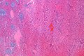
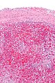
- Hyaline material adherent to splenic capsule.
- Hyaline material = pink acellular crap on a H&E stain.
Images
Hyaloserositis - intermed. mag. (WC)
Hyaloserositis - high mag. (WC)


Mononucleosis
General
- EBV infection.
- +/-Massive splenic enlargement.
Clinical:
- Monospot test +ve.
Microscopic
Features:
- Atypical lymphoid cells.
- Abundant basophilic cytoplasm.
- Cells indented by adjacent RBCs on blood smears.[11]
Images:
Flow cytometry
- CD8 >>> CD4.[12]
Littoral cell angioma
- For angioma see vascular malformations.
Splenic hamartoma
Splenic infarct
- AKA splenic infarction.
- AKA infarction of the spleen.
General
Classic textbook causes:[13]
- Septic embolus due to bacterial endocarditis.
- Sickle cell disease.
Usual causes:[13]
- Hematologic malignancy.
- Intracardiac thrombus.
- Bacterial endocarditis.
Clinical:[13]
- Left upper quadrant pain ~ 1/3 of cases.
- Fever ~ 1/3 of cases.
- Leukocytosis ~ 1/2 of cases.
Gross
- Classically wedge-shaped; triangular on section.
- The base of the triangle runs along the surface.
- The apex points to the obstructed vessel that lead to the infarct.
Microscopic
- See necrosis.
Weird stuff
- Dendritic cell tumours.[8]
- Interdigitating dendritic cell tumour.
- Follicular dendritic cell tumour.
Follicular dendritic cell tumour
- AKA follicular dendritic cell sarcoma.
Hepatosplenic T-cell lymphoma
- Abbreviated HSTL.
General
- Rare.
- Prognosis: poor.
Subtypes:[14]
- Hepatosplenic γδ T-cell lymphoma.
- May be written Hepatosplenic gamma/delta T-cell lymphoma.
- Hepatosplenic αβ T-cell lymphoma.
- May be written Hepatosplenic alpha/beta T-cell lymphoma.
Clinical triad:
- Hepatosplenomegaly.
- Cytopenias (anemia, thrombocytopenia).
- Sinusoidal tropism.
Microscopic
- Small cell lymphoma/Intermediate cell lymphoma.
- +/-"Folded" nuclei.
DDx:
Images:
IHC
Features:[9]
- CD4 -ve.
- CD8 -ve.
- NK cell-associated antigens +ve:[15]
- Effector proteins +ve.[15]
- Perforin, granzyme B, TIA-1, Fas ligand.
Myelolipoma of the spleen
Immune thrombocytopenic purpura
Spleens are removed for this.
Splenogonadal fusion
See also
References
- ↑ Humphrey, Peter A; Dehner, Louis P; Pfeifer, John D (2008). The Washington Manual of Surgical Pathology (1st ed.). Lippincott Williams & Wilkins. pp. 589. ISBN 978-0781765275.
- ↑ Aubrey-Bassler, FK.; Sowers, N. (Aug 2012). "613 cases of splenic rupture without risk factors or previously diagnosed disease: a systematic review.". BMC Emerg Med 12 (1): 11. doi:10.1186/1471-227X-12-11. PMID 22889306.
- ↑ Klatt, Edward C. (2006). Robbins and Cotran Atlas of Pathology (1st ed.). Saunders. pp. 91. ISBN 978-1416002741.
- ↑ Maakaroun, NR.; Moanna, A.; Jacob, JT.; Albrecht, H. (Mar 2010). "Viral infections associated with haemophagocytic syndrome.". Rev Med Virol 20 (2): 93-105. doi:10.1002/rmv.638. PMID 20127750.
- ↑ Shimizu-Kohno, K.; Kimura, Y.; Kiyasu, J.; Miyoshi, H.; Yoshida, M.; Ichikawa, R.; Niino, D.; Ohshima, K. (Sep 2012). "Malignant lymphoma of the spleen in Japan: a clinicopathological analysis of 115 cases.". Pathol Int 62 (9): 577-82. doi:10.1111/j.1440-1827.2012.02844.x. PMID 22924843.
- ↑ Bennett, M.; Schechter, GP. (Apr 2010). "Treatment of splenic marginal zone lymphoma: splenectomy versus rituximab.". Semin Hematol 47 (2): 143-7. doi:10.1053/j.seminhematol.2010.01.004. PMID 20350661.
- ↑ Baseggio L, Traverse-Glehen A, Callet-Bauchu E, et al. (March 2011). "Relevance of a scoring system including CD11c expression in the identification of splenic diffuse red pulp small B-cell lymphoma (SRPL)". Hematol Oncol 29 (1): 47–51. doi:10.1002/hon.957. PMID 20677173.
- ↑ 8.0 8.1 Humphrey, Peter A; Dehner, Louis P; Pfeifer, John D (2008). The Washington Manual of Surgical Pathology (1st ed.). Lippincott Williams & Wilkins. pp. 596. ISBN 978-0781765275.
- ↑ 9.0 9.1 9.2 Humphrey, Peter A; Dehner, Louis P; Pfeifer, John D (2008). The Washington Manual of Surgical Pathology (1st ed.). Lippincott Williams & Wilkins. pp. 595. ISBN 978-0781765275.
- ↑ URL: http://www.drugs.com/dict/sugar-coated-spleen.html. Accessed on: 1 September 2010.
- ↑ 11.0 11.1 URL: http://library.med.utah.edu/WebPath/EXAM/IMGQUIZ/hpfrm.html. Accessed on: 4 December 2011.
- ↑ URL: http://path.upmc.edu/cases/case37/gross.html. Accessed on: 2 January 2012.
- ↑ 13.0 13.1 13.2 Lawrence, YR.; Pokroy, R.; Berlowitz, D.; Aharoni, D.; Hain, D.; Breuer, GS.; Osler, W. (Jun 2010). "Splenic infarction: an update on William Osler's observations.". Isr Med Assoc J 12 (6): 362-5. PMID 20928991.
- ↑ 14.0 14.1 URL: http://www.webpathology.com/image.asp?case=378&n=15. Accessed on: 22 December 2011.
- ↑ 15.0 15.1 Salhany, KE.; Feldman, M.; Kahn, MJ.; Peritt, D.; Schretzenmair, RD.; Wilson, DM.; DiPaola, RS.; Glick, AD. et al. (Jun 1997). "Hepatosplenic gammadelta T-cell lymphoma: ultrastructural, immunophenotypic, and functional evidence for cytotoxic T lymphocyte differentiation.". Hum Pathol 28 (6): 674-85. PMID 9191001.
- ↑ Niitsu, N.; Kohri, M.; Togano, T.; Nakamine, H.; Nakamura, S.; Iwabuchi, K.; Higashihara, M. (Nov 2004). "Development of hepatosplenic gammadelta T-cell lymphoma with pancytopenia during early pregnancy: a case report and review of the literature.". Eur J Haematol 73 (5): 367-71. doi:10.1111/j.1600-0609.2004.00300.x. PMID 15458516.