Pulmonary infarct
Jump to navigation
Jump to search
| Pulmonary infarct | |
|---|---|
| Diagnosis in short | |
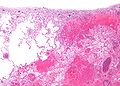
 Pulmonary infarct. H&E stain. | |
|
| |
| Synonyms | lung infarct |
|
| |
| LM | necrosis of alveolar walls - loss of nuclei, alveolar hemorrhage, +/-evidence of underlying cause |
| LM DDx | see Associated Dx |
| Gross | lung periphery, classically described as wedge-shaped |
| Site | lung |
|
| |
| Associated Dx | underlying causes: sickle cell disease, pulmonary embolism, vasculitides, malignancy (e.g. lymphoma), drug toxicity, others |
| Prevalence | uncommon |
| Radiology | reverse halo sign |
| Prognosis | dependent on underlying cause |
| Treatment | dependent on underlying cause |
Pulmonary infarct is the death of lung tissue due to oxygen deprivation.
It is also known as a lung infarct, lung infarction, and pulmonary infarction.
General
- Uncommon because of the dual blood supply (systemic via the bronchial arteries, pulmonary via the pulmonary arteries).
Common causes:[1]
Less common causes:
- Lymphoma, esp. acute promyelocytic leukemia.
- Drugs, e.g. chemotherapy.
- Vasculitis.
- Others.
Gross
- Lung periphery, classically described as wedge-shaped.
Note:
- In a histologic section, the classic wedge-shaped infarct is triangular:
- Base of triangle on the pleural aspect.
- Point furthest from the pleura close to the compromised artery that lead to infarction.
Radiology:
- Reverse halo sign.[2]
Images:
Microscopic
Features:
- Necrosis of alveolar walls - loss of nuclei.
- Alveolar hemorrhage.
Image
Pulmonary infarct - low mag. (WC)
See also
References
- ↑ URL: http://emedicine.medscape.com/article/908045-overview. Accessed on: 12 April 2012.
- ↑ 2.0 2.1 Casullo, J.; Semionov, A. (Feb 2013). "Reversed halo sign in acute pulmonary embolism and infarction.". Acta Radiol. doi:10.1177/0284185113475797. PMID 23395814.