Difference between revisions of "Pilar sheath acanthoma"
Jump to navigation
Jump to search
| (7 intermediate revisions by the same user not shown) | |||
| Line 3: | Line 3: | ||
| Image = SkinTumors-P8190634.JPG | | Image = SkinTumors-P8190634.JPG | ||
| Width = | | Width = | ||
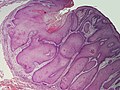
| Caption = | | Caption = Possibly a pilar sheath acanthoma. [[H&E stain]]. | ||
| Micro = | | Micro = cystic cavity with branching - contains keratineous material, usually extends from surface to deep cutis, cyst lining consists of a thickened squamous epithelium | ||
| Subtypes = | | Subtypes = | ||
| LMDDx = [[trichofolliculoma]], [[dilated pore of Winer]], [[epidermal inclusion cyst]] | | LMDDx = [[trichofolliculoma]], [[dilated pore of Winer]], [[epidermal inclusion cyst]] | ||
| Line 45: | Line 45: | ||
==Microscopic== | ==Microscopic== | ||
Features:<ref name=pmid718186/> | Features:<ref name=pmid718186/> | ||
*Cystic cavity with: | *Cystic cavity with branching:<ref name=pmid2697113>{{Cite journal | last1 = Choi | first1 = YS. | last2 = Park | first2 = SH. | last3 = Bang | first3 = D. | title = Pilar sheath acanthoma--report of a case with review of the literature. | journal = Yonsei Med J | volume = 30 | issue = 4 | pages = 392-5 | month = Dec | year = 1989 | doi = | PMID = 2697113 | URL = http://www.eymj.org/Synapse/Data/PDFData/0069YMJ/ymj-30-392.pdf }}</ref> | ||
** | **Contains keratineous material. | ||
**Usually extends from surface to deep cutis - like a hair shaft. | **Usually extends from surface to deep cutis - like a hair shaft. | ||
** | **Cyst lining consists of a thickened squamous epithelium ([[acanthosis]]). | ||
*+/-Mitoses. | |||
DDx: | DDx: | ||
*[[Trichofolliculoma]]. | *[[Trichofolliculoma]] - no branching of cavity, associated with hair & sebaceous glands.<ref name=pmid2697113/> | ||
*[[Dilated pore of Winer]]. | *[[Dilated pore of Winer]] - associated with hair & sebaceous glands.<ref name=pmid2697113/> | ||
*[[Epidermal inclusion cyst]]. | *[[Epidermal inclusion cyst]] - no branching, no accessory structures. | ||
===Images=== | ===Images=== | ||
<gallery> | <gallery> | ||
Image:SkinTumors-P8190634.JPG | | Image:SkinTumors-P8190634.JPG | Possibly a pilar sheath acanthoma. (WC) | ||
</gallery> | </gallery> | ||
www: | www: | ||
*[http://www.ncbi.nlm.nih.gov/pmc/articles/PMC3129125/figure/F1/ | *[http://www.ncbi.nlm.nih.gov/pmc/articles/PMC3129125/figure/F1/ Pilar sheath acanthoma (nih.gov)].<ref name=pmid21769237>{{Cite journal | last1 = Bavikar | first1 = RR. | last2 = Gaopande | first2 = V. | last3 = Deshmukh | first3 = SD. | title = Postauricular pilar sheath acanthoma. | journal = Int J Trichology | volume = 3 | issue = 1 | pages = 39-40 | month = Jan | year = 2011 | doi = 10.4103/0974-7753.82136 | PMID = 21769237 }}</ref> | ||
*[http://www.dermnet.org.nz/pathology/pilar-sheath-path.html Pilar sheath acanthoma (dermnet.org.nz)]. | *[http://www.dermnet.org.nz/pathology/pilar-sheath-path.html Pilar sheath acanthoma - several images (dermnet.org.nz)]. | ||
*[http://www.eymj.org/Synapse/Data/PDFData/0069YMJ/ymj-30-392.pdf Pilar sheath acanthoma (eymj.org)].<ref name=pmid2697113/> | |||
*[http://www.dermatopathonline.com/pilar%20sheath%20acanthoma2.html Pilar sheath acanthoma - several images (dermatopathonline.com)]. | |||
==See also== | ==See also== | ||
*[[Dermatopathology]]. | *[[Dermatopathology]]. | ||
*[[Skin cysts]]. | *[[Skin cysts]]. | ||
*[[Non-malignant skin disease]]. | |||
==References== | ==References== | ||
Latest revision as of 12:36, 30 October 2013
| Pilar sheath acanthoma | |
|---|---|
| Diagnosis in short | |
 Possibly a pilar sheath acanthoma. H&E stain. | |
|
| |
| LM | cystic cavity with branching - contains keratineous material, usually extends from surface to deep cutis, cyst lining consists of a thickened squamous epithelium |
| LM DDx | trichofolliculoma, dilated pore of Winer, epidermal inclusion cyst |
| Gross | raised lesion, central cavitation |
| Site | skin - classically upper lip |
|
| |
| Clinical history | typically middle aged - 50s |
| Prevalence | rare |
| Prognosis | benign |
Pilar sheath acanthoma is a rare benign skin lesion.
General
- Rare.[1]
- Considered a hamartoma.[1]
- Does not regress like keratoacanthoma.
- Typically middle aged individuals - median age in one series 55 years old, range 46-75 years old.[2]
Gross
- Raised lesion with a central depression - somewhat similar to keratoacanthoma.
- Classcially upper lip.[2]
DDx:
Microscopic
Features:[2]
- Cystic cavity with branching:[3]
- Contains keratineous material.
- Usually extends from surface to deep cutis - like a hair shaft.
- Cyst lining consists of a thickened squamous epithelium (acanthosis).
- +/-Mitoses.
DDx:
- Trichofolliculoma - no branching of cavity, associated with hair & sebaceous glands.[3]
- Dilated pore of Winer - associated with hair & sebaceous glands.[3]
- Epidermal inclusion cyst - no branching, no accessory structures.
Images
Possibly a pilar sheath acanthoma. (WC)
www:
- Pilar sheath acanthoma (nih.gov).[4]
- Pilar sheath acanthoma - several images (dermnet.org.nz).
- Pilar sheath acanthoma (eymj.org).[3]
- Pilar sheath acanthoma - several images (dermatopathonline.com).
See also
References
- ↑ 1.0 1.1 Vakilzadeh, F. (Jan 1987). "[Pilar sheath acanthoma].". Hautarzt 38 (1): 40-2. PMID 3557980.
- ↑ 2.0 2.1 2.2 Mehregan, AH.; Brownstein, MH. (Oct 1978). "Pilar sheath acanthoma.". Arch Dermatol 114 (10): 1495-7. PMID 718186.
- ↑ 3.0 3.1 3.2 3.3 Choi, YS.; Park, SH.; Bang, D. (Dec 1989). "Pilar sheath acanthoma--report of a case with review of the literature.". Yonsei Med J 30 (4): 392-5. PMID 2697113.
- ↑ Bavikar, RR.; Gaopande, V.; Deshmukh, SD. (Jan 2011). "Postauricular pilar sheath acanthoma.". Int J Trichology 3 (1): 39-40. doi:10.4103/0974-7753.82136. PMID 21769237.