Difference between revisions of "Pericardium"
Jump to navigation
Jump to search
| (7 intermediate revisions by the same user not shown) | |||
| Line 1: | Line 1: | ||
[[Image:Blausen 0470 HeartWall.png|thumb|right|300px|The heart wall (schematic). (WC/Wikiversity Journal of Medicine)]] | |||
'''Pericardium''' is a specimen that uncommonly comes to [[pathology]]. | '''Pericardium''' is a specimen that uncommonly comes to [[pathology]]. | ||
| Line 6: | Line 7: | ||
*Infectious pericarditis - post-procedural. | *Infectious pericarditis - post-procedural. | ||
*[[Fibrinous pericarditis]]. | *[[Fibrinous pericarditis]]. | ||
*Pericardial cyst. | *[[Pericardial cyst]]. | ||
===Malignant=== | ===Malignant=== | ||
| Line 21: | Line 20: | ||
=Specific entities= | =Specific entities= | ||
==Idiopathic pericarditis== | ==Idiopathic pericarditis== | ||
:''Pericarditis'' redirects here. | |||
===General=== | ===General=== | ||
*Uncommon. | *Uncommon. | ||
*In the clinical context ''pericarditis'' is used for things that probably don't have inflammation.<ref name=pmid16200146>{{Cite journal | last1 = Roberts | first1 = WC. | title = Pericardial heart disease: its morphologic features and its causes. | journal = Proc (Bayl Univ Med Cent) | volume = 18 | issue = 1 | pages = 38-55 | month = Jan | year = 2005 | doi = | PMID = 16200146 }}</ref> | |||
**"Pericardial heart disease" may be a better descriptor. | |||
Etiologies of pericarditis:<ref name=pmid22723538/> | Etiologies of pericarditis:<ref name=pmid22723538/> | ||
| Line 55: | Line 57: | ||
*[[Malignant mesothelioma]].<ref name=pmid23773902/> | *[[Malignant mesothelioma]].<ref name=pmid23773902/> | ||
==Sign out== | ====Images==== | ||
<gallery> | |||
Image: Acute pericarditis -- intermed mag.jpg | AP - intermed. mag. (WC) | |||
Image: Acute pericarditis -- high mag.jpg | AP - high mag. (WC) | |||
Image: Acute pericarditis -- very high mag.jpg | AP - very high mag. (WC) | |||
</gallery> | |||
===Sign out=== | |||
<pre> | <pre> | ||
PERICARDIUM, BIOPSY: | PERICARDIUM, BIOPSY: | ||
- CHRONIC PERICARDITIS WITH SIDEROPHAGES AND REACTIVE MESOTHELIAL CHANGES. | - ACUTE AND CHRONIC PERICARDITIS WITH SIDEROPHAGES AND REACTIVE MESOTHELIAL CHANGES. | ||
- NO MICRO-ORGANISMS SEEN WITH ROUTINE STAINING. | |||
- NO EVIDENCE OF MALIGNANCY. | - NO EVIDENCE OF MALIGNANCY. | ||
</pre> | </pre> | ||
===Micro=== | ====Micro==== | ||
The sections show fibrous tissue with minimal adipose tissue that is covered by mesothelium. A mixed inflammatory infiltrate is present that consists primarily of lymphocytes and plasma cells. Rare eosinophils are seen. Abundant hemosideratin-laden macrophages are seen. | The sections show fibrous tissue with minimal adipose tissue that is covered by mesothelium. A mixed inflammatory infiltrate is present that consists primarily of lymphocytes and plasma cells. Rare eosinophils are seen. Focally, neutrophils are seen and associated with reactive mesothelial cells. Abundant hemosideratin-laden macrophages are seen. No fibrinous strands are seen. No significant nuclear atypia is identified and no atypical infiltrative cell population is identified. No micro-organisms are identified with routine staining. | ||
==See also== | ==See also== | ||
*[[Heart]]. | *[[Heart]]. | ||
*[[Mediastinum]]. | *[[Mediastinum]]. | ||
*[[Fibrinous pericarditis]]. | |||
==References== | ==References== | ||
Latest revision as of 12:12, 8 July 2016

Pericardium is a specimen that uncommonly comes to pathology.
Pathologies of the pericardium
Benign
- Idiopathic pericarditis - common.[1]
- Infectious pericarditis - post-procedural.
- Fibrinous pericarditis.
- Pericardial cyst.
Malignant
Related pathologies
- Pericardial effusion.
- Hemopericardium.
- Cardiac tamponade.
Specific entities
Idiopathic pericarditis
- Pericarditis redirects here.
General
- Uncommon.
- In the clinical context pericarditis is used for things that probably don't have inflammation.[2]
- "Pericardial heart disease" may be a better descriptor.
Etiologies of pericarditis:[3]
- Infectious:
- Fungal.
- Bacterial.
- Idiopathic - most common.
- Neoplastic.
- Autoimmune, e.g. systemic lupus erythematosus.
- Uremia - chronic renal failure.
- Traumatic - post-surgical.
- Associated with myocardial infarction - Dressler's syndrome.
Gross
Features:[3]
- Thickening.
- +/-Pericardial effusion.
Notes:
- Normal pericardial fluid volume 5-35 mL.[3]
Microscopic
Features:
- Inflammatory cells:
- Neutrophils.
- Lymphocytes.
- Plasma cells.
- +/-Hemosiderin-laden macrophages.
DDx:
- Infectious pericarditis.
- Malignant mesothelioma.[1]
Images
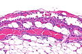
AP - intermed. mag. (WC)
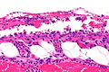
AP - high mag. (WC)
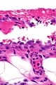
AP - very high mag. (WC)


Sign out
PERICARDIUM, BIOPSY:
- ACUTE AND CHRONIC PERICARDITIS WITH SIDEROPHAGES AND REACTIVE MESOTHELIAL CHANGES.
- NO MICRO-ORGANISMS SEEN WITH ROUTINE STAINING.
- NO EVIDENCE OF MALIGNANCY.
Micro
The sections show fibrous tissue with minimal adipose tissue that is covered by mesothelium. A mixed inflammatory infiltrate is present that consists primarily of lymphocytes and plasma cells. Rare eosinophils are seen. Focally, neutrophils are seen and associated with reactive mesothelial cells. Abundant hemosideratin-laden macrophages are seen. No fibrinous strands are seen. No significant nuclear atypia is identified and no atypical infiltrative cell population is identified. No micro-organisms are identified with routine staining.
See also
References
- ↑ 1.0 1.1 Smets P, Guettrot-Imbert G, Hermet M, et al. (September 2013). "[Recurrent pericarditis related to primary pericardial malignant mesothelioma]" (in French). Rev Med Interne 34 (9): 573–6. doi:10.1016/j.revmed.2013.04.021. PMID 23773902.
- ↑ Roberts, WC. (Jan 2005). "Pericardial heart disease: its morphologic features and its causes.". Proc (Bayl Univ Med Cent) 18 (1): 38-55. PMID 16200146.
- ↑ 3.0 3.1 3.2 Peebles CR, Shambrook JS, Harden SP (December 2011). "Pericardial disease--anatomy and function". Br J Radiol 84 Spec No 3: S324–37. doi:10.1259/bjr/16168253. PMC 3473919. PMID 22723538. https://www.ncbi.nlm.nih.gov/pmc/articles/PMC3473919/.