Partial colectomy for diverticular disease
Jump to navigation
Jump to search
Partial colectomy for diverticular disease is a very common procedure. The sigmoid colon is typically afflicited; in that case it can more precisely be labeled sigmoidectomy for diverticular disease.
Introduction
This is quite common. Diverticulitis (inflammation of diverticula) and it complications are usually diagnosed by medical imaging.
If the individual has a peritonitis, a (temporary) stoma is generally created.
The pathologist's main tasks in this specimen is:
- Confirming and documenting extent of the disease.
- Excluding malignancy.
Protocol
Specimen:
- Length __ cm.
- Circumference (proximal/one end) __ cm.
- Circumference (distal/other end) __ cm.
- Mesentry (maximal): __ cm.
Appearance:
- Serosal surface: [shiny/hemorrhagic/dull/exudate/adhesions].
- Mucosa: [unremarkable/granular].
- Polyps: [none/number - size __ cm, location (to nearest resection margin): __ cm].
- Number of diverticula (count up to 6, then estimate): [number of diverticula].
- Wall: [unremarkable/thickened].
Other:
- Perforation: [not identified/present - location of perforation (to nearest mucosal margin): __ cm, size of performation __ cm].
Representative sections submitted:
- Proximal mucosal margin.
- Distal mucosal margin.
- Diverticula (3-5 blocks).
- Interdiverticular mucosa (2 blocks).
- Lymph nodes (2 blocks).
Protocol notes
Alternate approaches
Pathology
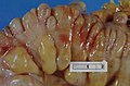
Diverticular disease.
