Difference between revisions of "Neuroblastoma"
m (redirect for now) |
Jensflorian (talk | contribs) (→Classification/grading: INRGSS) |
||
| (4 intermediate revisions by 2 users not shown) | |||
| Line 1: | Line 1: | ||
'''Neuroblastoma''' is a [[malignant]] tumour of the [[adrenal gland]]. | |||
''[[Olfactory neuroblastoma]]'' is dealt with separately. | |||
==General== | |||
Epidemiology: | |||
*Usually paediatric population. | |||
Laboratory findings: | |||
*Increased urine homovanillic acid. | |||
Imaging: | |||
*mIBG uptake (>90% neuroblastomas) | |||
Predictors of a poor prognosis:<ref name=Ref_PCPBoD8_254>{{Ref PCPBoD8|254}}</ref> | |||
*High mitotic-karyorrhectic index. | |||
*Lack of schwannian stroma. | |||
*>18 months. | |||
*Near ploidy. | |||
*N-MYC amplification. | |||
*1p deletion /imbalance | |||
*Lymph node spread. | |||
*Distant spread. | |||
Classification: | |||
*In a grouping known as ''neuroblastic tumours'' which includes:<ref name=pmid10421272>{{cite journal |author=Shimada H, Ambros IM, Dehner LP, Hata J, Joshi VV, Roald B |title=Terminology and morphologic criteria of neuroblastic tumors: recommendations by the International Neuroblastoma Pathology Committee |journal=Cancer |volume=86 |issue=2 |pages=349–63 |year=1999 |month=July |pmid=10421272 |doi= |url=}}</ref> | |||
**[[Ganglioneuroma]] (benign). | |||
**[[Ganglioneuroblastoma]] (intermediate). | |||
**Neuroblastoma (aggressive). | |||
==Gross== | |||
*Typically an abdominal mass. | |||
**~40% arise in the [[adrenal gland]].<ref name=Ref_PCPBoD8_253>{{Ref PCPBoD8|253}}</ref> | |||
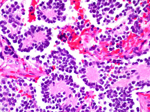
==Microscopic== | |||
Features:<ref name=pmid18635637>{{cite journal |author=Chung EM, Murphey MD, Specht CS, Cube R, Smirniotopoulos JG |title=From the Archives of the AFIP. Pediatric orbit tumors and tumorlike lesions: osseous lesions of the orbit |journal=Radiographics |volume=28 |issue=4 |pages=1193–214 |year=2008 |pmid=18635637 |doi=10.1148/rg.284085013 |url=}}</ref> | |||
*[[small round cell tumour|Small round blue cell]]s separated by thin (pink) fibrous septa. | |||
*Homer-Wright rosettes. | |||
**Rosette with a small (~100 micrometers - diameter) meshwork of fibers (neuropil) at the centre.<ref name=pmid16551982>{{cite journal |author=Wippold FJ, Perry A |title=Neuropathology for the neuroradiologist: rosettes and pseudorosettes |journal=AJNR Am J Neuroradiol |volume=27 |issue=3 |pages=488–92 |year=2006 |month=March |pmid=16551982 |doi= |url=}}</ref> | |||
*Neuropil-like stroma = paucicellular stroma with a cotton candy-like appearance; see comparison below. | |||
**>50% neuropil-like stroma -- otherwise it's a [[ganglioneurona]] or ganglioblastoma. | |||
Notes: | |||
*The fibrous septa are especially useful for differentiation from lymphoma. | |||
DDx: | |||
*[[Small round cell tumours]]. | |||
**[[Wilms tumour]]. | |||
**Lymphoma. | |||
**[[Hepatoblastoma]]. | |||
===Images=== | |||
<gallery> | |||
Image:Adrenal Neuroblastoma MP CTR.jpg|Neuroblastoma - medium power]] | |||
Image:Adrenal Neuroblastoma 2 MP CTR.jpg|Neuroblastoma - medium power (SKB) | |||
Image:Adrenal Neuroblastoma M2P PA.JPG|Neuroblastoma - medium power]] | |||
Image:Adrenal Neuroblastoma VascularInvasion MP CTR.jpg|thumb|Neuroblastoma - vascular invasion - medium power]] | |||
Image:Adrenal Neuroblastoma MP3 PA.JPG|Neuroblastoma - medium power]] | |||
Image:Adrenal Neuroblastoma MP PA.JPG|Neuroblastoma - medium power]] | |||
Image:Adrenal Neuroblastoma 2 HP CTR.jpg|Neuroblastoma - high power (SKB) | |||
Image:Adrenal Neuroblastoma 3 HP CTR.jpg|Neuroblastoma - high power - blue cells arrayed around a core of fluffy pink neuropil (SKB) | |||
Image:Adrenal Neuroblastoma HP2 CTR.jpg|Neuroblastoma - high power]] | |||
Image:Adrenal Neuroblastoma HP CTR.jpg|Neuroblastoma - high power]] | |||
</gallery> | |||
*[http://radiographics.rsna.org/content/28/4/1193/F42.expansion Neuroblastoma (radiographics.rsna.org)].<ref>URL: [http://radiographics.rsna.org/content/28/4/1193.full http://radiographics.rsna.org/content/28/4/1193.full]. Accessed on: 12 January 2011.</ref> | |||
*[http://commons.wikimedia.org/wiki/File:Neuroblastoma_rosettes.jpg Neuroblastoma (WC)]. | |||
*[http://farm3.static.flickr.com/2259/2274260465_afbea05f9b.jpg Neuroblastoma (flickr.com)]. | |||
===Schwannian vs. neuropil=== | |||
{| class="wikitable sortable" style="margin-left:auto;margin-right:auto" | |||
| Feature | |||
| Schwannian | |||
| Neuropil | |||
|- | |||
| Cellularity | |||
| high ~ spacing of cells < 30 µm | |||
| low ~ spacing of cells > 100 µm | |||
|- | |||
| Fibrillary | |||
| yes, long fine strands | |||
| no | |||
|- | |||
| Associations | |||
| ganglion cells | |||
| neuroblasts | |||
|- | |||
| Cytoplasmic vacuolation | |||
| yes | |||
| ? | |||
|- | |||
|} | |||
===Classification/grading=== | |||
Commonly grouped by the ''Shimada classification'', which depends on the presence a number of things including: | |||
*Mitoses/karyorrhectic cells. | |||
*Molecular abnormalities. | |||
International Neuroblastoma Risk Group Staging System (INRGSS) | |||
* L1: Locoregional tumor without Image-Definging Risk factors (IDRFs) | |||
* L2: Locoregional tumor with one or more IDRFs | |||
* M: Distant metastatic disease (except Ms) | |||
* Ms: INRG Stage L1 or L2 tumor with metastatic disease confined to skin and/or liver and/or bone marrow and age below 18 months. | |||
==IHC== | |||
*PGP 9.5 +ve.<ref>{{Cite journal | last1 = Ootsuka | first1 = S. | last2 = Asami | first2 = S. | last3 = Sasaki | first3 = T. | last4 = Yoshida | first4 = Y. | last5 = Nemoto | first5 = N. | last6 = Shichino | first6 = H. | last7 = Chin | first7 = M. | last8 = Mugishima | first8 = H. | last9 = Suzuki | first9 = T. | title = Useful markers for detecting minimal residual disease in cases of neuroblastoma. | journal = Biol Pharm Bull | volume = 31 | issue = 6 | pages = 1071-4 | month = Jun | year = 2008 | doi = | PMID = 18520032 }}</ref> | |||
**PGP = protein gene product. | |||
*NB-84 +ve.<ref name=pmid9500774>{{Cite journal | last1 = Miettinen | first1 = M. | last2 = Chatten | first2 = J. | last3 = Paetau | first3 = A. | last4 = Stevenson | first4 = A. | title = Monoclonal antibody NB84 in the differential diagnosis of neuroblastoma and other small round cell tumors. | journal = Am J Surg Pathol | volume = 22 | issue = 3 | pages = 327-32 | month = Mar | year = 1998 | doi = | PMID = 9500774 }}</ref> | |||
**More sensitive that synaptophysin. | |||
*Synaptophysin +ve. | |||
*CD99 -ve. | |||
==EM== | |||
Distinctive EM appearance:<ref name=pmid1196755>{{Cite journal | last1 = Mackay | first1 = B. | last2 = Masse | first2 = SR. | last3 = King | first3 = OY. | last4 = Butler | first4 = J. | title = Diagnosis of neuroblastoma by electron microscopy of bone marrow aspirates. | journal = Pediatrics | volume = 56 | issue = 6 | pages = 1045-9 | month = Dec | year = 1975 | doi = | PMID = 1196755 }}</ref> | |||
*Dendritic processes with longitudinally oriented microtubules. | |||
*Membrane bound electron-dense granules (contain catecholamines). | |||
*Desmosomes | |||
**Not seen in [[EWS]], [[RMS]], lymphomas. | |||
*Membrane densities. | |||
Pertinent negative:<ref name=pmid1196755/> | |||
*No glycogen. | |||
**Seen in [[EWS]]. | |||
==See also== | |||
*[[Adrenal gland]]. | |||
==References== | |||
{{Reflist|1}} | |||
[[Category:Diagnosis]] | |||
[[Category:Adrenal gland]] | |||
Latest revision as of 09:40, 7 March 2019
Neuroblastoma is a malignant tumour of the adrenal gland.
Olfactory neuroblastoma is dealt with separately.
General
Epidemiology:
- Usually paediatric population.
Laboratory findings:
- Increased urine homovanillic acid.
Imaging:
- mIBG uptake (>90% neuroblastomas)
Predictors of a poor prognosis:[1]
- High mitotic-karyorrhectic index.
- Lack of schwannian stroma.
- >18 months.
- Near ploidy.
- N-MYC amplification.
- 1p deletion /imbalance
- Lymph node spread.
- Distant spread.
Classification:
- In a grouping known as neuroblastic tumours which includes:[2]
- Ganglioneuroma (benign).
- Ganglioneuroblastoma (intermediate).
- Neuroblastoma (aggressive).
Gross
- Typically an abdominal mass.
- ~40% arise in the adrenal gland.[3]
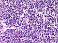
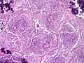
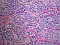
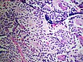
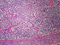
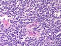
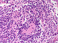
Microscopic
Features:[4]
- Small round blue cells separated by thin (pink) fibrous septa.
- Homer-Wright rosettes.
- Rosette with a small (~100 micrometers - diameter) meshwork of fibers (neuropil) at the centre.[5]
- Neuropil-like stroma = paucicellular stroma with a cotton candy-like appearance; see comparison below.
- >50% neuropil-like stroma -- otherwise it's a ganglioneurona or ganglioblastoma.
Notes:
- The fibrous septa are especially useful for differentiation from lymphoma.
DDx:
- Small round cell tumours.
- Wilms tumour.
- Lymphoma.
- Hepatoblastoma.
Images
Neuroblastoma - medium power]]
Neuroblastoma - medium power (SKB)
Neuroblastoma - medium power]]
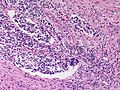
Neuroblastoma - vascular invasion - medium power]]
Neuroblastoma - medium power]]
Neuroblastoma - medium power]]
Neuroblastoma - high power (SKB)
Neuroblastoma - high power - blue cells arrayed around a core of fluffy pink neuropil (SKB)
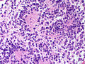
Neuroblastoma - high power]]
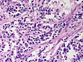
Neuroblastoma - high power]]


Schwannian vs. neuropil
| Feature | Schwannian | Neuropil |
| Cellularity | high ~ spacing of cells < 30 µm | low ~ spacing of cells > 100 µm |
| Fibrillary | yes, long fine strands | no |
| Associations | ganglion cells | neuroblasts |
| Cytoplasmic vacuolation | yes | ? |
Classification/grading
Commonly grouped by the Shimada classification, which depends on the presence a number of things including:
- Mitoses/karyorrhectic cells.
- Molecular abnormalities.
International Neuroblastoma Risk Group Staging System (INRGSS)
- L1: Locoregional tumor without Image-Definging Risk factors (IDRFs)
- L2: Locoregional tumor with one or more IDRFs
- M: Distant metastatic disease (except Ms)
- Ms: INRG Stage L1 or L2 tumor with metastatic disease confined to skin and/or liver and/or bone marrow and age below 18 months.
IHC
- PGP 9.5 +ve.[7]
- PGP = protein gene product.
- NB-84 +ve.[8]
- More sensitive that synaptophysin.
- Synaptophysin +ve.
- CD99 -ve.
EM
Distinctive EM appearance:[9]
- Dendritic processes with longitudinally oriented microtubules.
- Membrane bound electron-dense granules (contain catecholamines).
- Desmosomes
- Membrane densities.
Pertinent negative:[9]
- No glycogen.
- Seen in EWS.
See also
References
- ↑ Mitchell, Richard; Kumar, Vinay; Fausto, Nelson; Abbas, Abul K.; Aster, Jon (2011). Pocket Companion to Robbins & Cotran Pathologic Basis of Disease (8th ed.). Elsevier Saunders. pp. 254. ISBN 978-1416054542.
- ↑ Shimada H, Ambros IM, Dehner LP, Hata J, Joshi VV, Roald B (July 1999). "Terminology and morphologic criteria of neuroblastic tumors: recommendations by the International Neuroblastoma Pathology Committee". Cancer 86 (2): 349–63. PMID 10421272.
- ↑ Mitchell, Richard; Kumar, Vinay; Fausto, Nelson; Abbas, Abul K.; Aster, Jon (2011). Pocket Companion to Robbins & Cotran Pathologic Basis of Disease (8th ed.). Elsevier Saunders. pp. 253. ISBN 978-1416054542.
- ↑ Chung EM, Murphey MD, Specht CS, Cube R, Smirniotopoulos JG (2008). "From the Archives of the AFIP. Pediatric orbit tumors and tumorlike lesions: osseous lesions of the orbit". Radiographics 28 (4): 1193–214. doi:10.1148/rg.284085013. PMID 18635637.
- ↑ Wippold FJ, Perry A (March 2006). "Neuropathology for the neuroradiologist: rosettes and pseudorosettes". AJNR Am J Neuroradiol 27 (3): 488–92. PMID 16551982.
- ↑ URL: http://radiographics.rsna.org/content/28/4/1193.full. Accessed on: 12 January 2011.
- ↑ Ootsuka, S.; Asami, S.; Sasaki, T.; Yoshida, Y.; Nemoto, N.; Shichino, H.; Chin, M.; Mugishima, H. et al. (Jun 2008). "Useful markers for detecting minimal residual disease in cases of neuroblastoma.". Biol Pharm Bull 31 (6): 1071-4. PMID 18520032.
- ↑ Miettinen, M.; Chatten, J.; Paetau, A.; Stevenson, A. (Mar 1998). "Monoclonal antibody NB84 in the differential diagnosis of neuroblastoma and other small round cell tumors.". Am J Surg Pathol 22 (3): 327-32. PMID 9500774.
- ↑ 9.0 9.1 Mackay, B.; Masse, SR.; King, OY.; Butler, J. (Dec 1975). "Diagnosis of neuroblastoma by electron microscopy of bone marrow aspirates.". Pediatrics 56 (6): 1045-9. PMID 1196755.