Difference between revisions of "Necrobiosis lipoidica"
Jump to navigation
Jump to search
(tweak) |
|||
| Line 14: | Line 14: | ||
| Gross = | | Gross = | ||
| Grossing = | | Grossing = | ||
| Site = | | Site = [[skin]] | ||
| Assdx = [[diabetes mellitus]], [[rheumatoid arthritis]] | | Assdx = [[diabetes mellitus]], [[rheumatoid arthritis]] | ||
| Syndromes = | | Syndromes = | ||
Revision as of 01:53, 31 October 2013
| Necrobiosis lipoidica | |
|---|---|
| Diagnosis in short | |
 Necrobiosis lipoidica. HPS stain. | |
|
| |
| LM | (dermal) palisading granulomas around necrotic collagen; little mucin; no normal dermis between foci; plasma cells - common; +/-involvement of subcutis |
| LM DDx | granuloma annulare, rheumatoid nodule |
| Site | skin |
|
| |
| Associated Dx | diabetes mellitus, rheumatoid arthritis |
| Prevalence | uncommon |
Necrobiosis lipoidica is an uncommon granulomatous skin pathology.
General
Associated with:
- Diabetes mellitus - known as necrobiosis lipoidica diabeticorum.
- Rheumatoid arthritis.
Gross
Image:
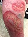
Necrobiosis lipoidica. (WC)

Microscopic
Features:[1]
- Dermal palisading granuloma around:
- Necrotic collagen - key feature.
- Nuclei "missing" - have undergone karyolysis.
- Necrotic collagen - key feature.
- Little mucin, no normal dermis between foci.
- Plasma cells - common.[2]
- May involve adipose tissue.
DDx:
- Granuloma annulare - more mucin, normal dermis between foci,[1] plasma cells uncommon,[2] no fat involvement - usu. more superficial.
- Rheumatoid nodule.
Images
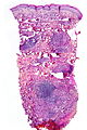
Necrobiosis lipoidica - low mag. (WC)
www:
- Necrobiosis lipoidica (dermatology.cdlib.org).
- Necrobiosis lipoidica (drmihm.com).
- Necrobiosis lipoidica (dermnetnz.org).
See also
- [Non-malignant skin disease]].
References
- ↑ 1.0 1.1 Busam, Klaus J. (2009). Dermatopathology: A Volume in the Foundations in Diagnostic Pathology Series (1st ed.). Saunders. pp. 51. ISBN 978-0443066542.
- ↑ 2.0 2.1 URL: http://dermnetnz.org/pathology/necrobiosis-lipoidica-path.html. Accessed on: 24 January 2012.