Myxopapillary ependymoma
| Myxopapillary ependymoma | |
|---|---|
| Diagnosis in short | |
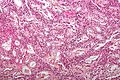
 Myxopapillary ependymoma. H&E stain | |
|
| |
| LM | Papillary tumor cells around vascular myxoid matrix |
| Subtypes | subtype of ependymoma |
| LM DDx | chordoma, myxoid chondrosarcoma, paraganglioma, papillary adenocarcinoma |
| Stains | Alcian blue +ve |
| IHC | GFAP +ve |
| Site | usually lumbar spinal cord |
|
| |
| Prognosis | good (WHO Grade I) |
Mxyopapillary Ependymoma, is a low-grade Ependymoma. It is nearly always associated with cauda equina and filum terminale.
General
- Low-grade ependymoma - WHO Grade I by definition.
- Classically in the spinal cord of adults.
- Approx 9-13% of all ependymal tumors.[1]
- Associated with back pain.
- Enhancing mass
Gross
- Soft.
- Gray.
- Discrete masses.
- Often encapsulated.
- Subtotal resected tumors may spread throughout the neuraxis.
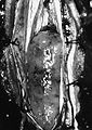
Myxopapillary ependymoma in the filum terminale (Dr. E. Michael Scott, Boston, MA).
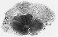
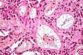
Mxyopapillary Ependymoma in the spinal cord, cross section (AFIP)


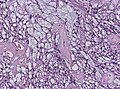
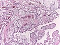
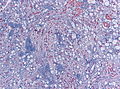
Microscopic
Features:
- Papillary appearance.
- Perivascular pseudorosettes:
- Cuboidal to elongated tumor cells.
- Radially arranged around vascular cores.
- Myxoid material surround blood vessels.
- Microcysts.
- Low mitotic activity.
Note: Cases with extensive sclerosis may mimic degenerative changes. [2]
DDx:
- Chordoma.
- Myxoid chondrosarcoma.
- Paraganglioma.
- Papillary adenocarcinoma.
Images
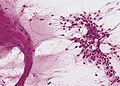
Mxyopapillary Ependymoma, HE Smear (AFIP)
Mxyopapillary Ependymoma, low magnification (WC/Nephron)
Intermediate magnification (WC/jensflorian)
High magnification (WC/Nephron)
Mxyopapillary Ependymoma, vascular sclerosis (WC/jensflorian)
Alcian-blue staining highlighting the mucinous matrix (WC/jensflorian)


- Myxopapillary ependymoma - high mag. (WC).
- Myxopapillary ependymoma (bmj.com) - part of careers.bmj.com article on paediatric pathology.
- Myxopapillary ependymoma - cytology (WC).
- Myxopapillary ependymoma - several images (upmc.edu).
IHC
- GFAP+ve.
- S-100+ve.
- MIB-1 <1%.
Molecular
- Poorly characterized.
- No consistent abberations.
See also
References
- ↑ Schiffer, D.; Chiò, A.; Giordana, MT.; Migheli, A.; Palma, L.; Pollo, B.; Soffietti, R.; Tribolo, A. (Aug 1991). "Histologic prognostic factors in ependymoma.". Childs Nerv Syst 7 (4): 177-82. PMID 1933913.
- ↑ Schittenhelm, J.; Becker, R.; Capper, D.; Meyermann, R.; Iglesias-Rozas, JR.; Kaminsky, J.; Mittelbronn, M.. "The clinico-surgico-pathological spectrum of myxopapillary ependymomas--report of four unusal cases and review of the literature.". Clin Neuropathol 27 (1): 21-8. PMID 18257471.