Difference between revisions of "Mesenchymal chondrosarcoma"
Jump to navigation
Jump to search
(split out) |
|||
| Line 37: | Line 37: | ||
*SOX9 (positive in small cells and chondrocytes) <ref>{{Cite journal | last1 = Pang | first1 = ZG. | last2 = He | first2 = XZ. | last3 = Wu | first3 = LY. | last4 = Wei | first4 = W. | last5 = Liu | first5 = XY. | last6 = Liao | first6 = DY. | last7 = Li | first7 = FY. | last8 = Zhang | first8 = XL. | title = [Clinicopathologic and immunohistochemical study of 23 cases of mesenchymal chondrosarcoma]. | journal = Zhonghua Bing Li Xue Za Zhi | volume = 40 | issue = 6 | pages = 368-72 | month = Jun | year = 2011 | doi = | PMID = 21914343 }}</ref> | *SOX9 (positive in small cells and chondrocytes) <ref>{{Cite journal | last1 = Pang | first1 = ZG. | last2 = He | first2 = XZ. | last3 = Wu | first3 = LY. | last4 = Wei | first4 = W. | last5 = Liu | first5 = XY. | last6 = Liao | first6 = DY. | last7 = Li | first7 = FY. | last8 = Zhang | first8 = XL. | title = [Clinicopathologic and immunohistochemical study of 23 cases of mesenchymal chondrosarcoma]. | journal = Zhonghua Bing Li Xue Za Zhi | volume = 40 | issue = 6 | pages = 368-72 | month = Jun | year = 2011 | doi = | PMID = 21914343 }}</ref> | ||
*S100 (positive in chondrocytes not in small cells) | *S100 (positive in chondrocytes not in small cells) | ||
*Osteocalcin (negative in small cells) | *Osteocalcin (negative in small cells) | ||
==Molecular== | ==Molecular== | ||
| Line 44: | Line 44: | ||
==Photos== | ==Photos== | ||
<gallery> | <gallery> | ||
Image:Bone Chondrosarcoma Mesenchymal MP5 PA.JPG|Islands of cartilage in a background of small blue cells.(SKB) | |||
Image:Bone Chondrosarcoma Mesenchymal MP PA.JPG|Islands of cartilage in a background of small blue cells.(SKB) | |||
Image:Bone Chondrosarcoma Mesenchymal LP2 PA.JPG|Islands of cartilage in a background of small blue cells.(SKB) | |||
Image:Bone Chondrosarcoma Mesenchymal HP5 PA.JPG|Islands of cartilage in a background of small blue cells.(SKB) | |||
Image:Bone Chondrosarcoma Mesenchymal MP2 PA.JPG|Islands of cartilage in a background of small blue cells.(SKB) | |||
Image:Bone Chondrosarcoma Mesenchymal MP7.JPG|Islands of cartilage in a background of small blue cells.(SKB) | |||
Image:Bone Chondrosarcoma Mesenchymal HP3 PA.JPG|Small blue cells predominate.(SKB) | |||
</gallery> | </gallery> | ||
Revision as of 09:07, 20 November 2014
Mesenchymal chondrosarcoma is a type chondrosarcoma.
- Arise in soft tissue; this is where the name comes from.[1]
- Rare variant of chondrosarcoma.
Microscopic: Features:
- "White clouds in a blue sky".
Image:
General
- Rare variant of chondrosarcoma.
- 2–10% of primary chondrosarcomas
- Adolescents and young adults
- Female predilection
- Most commonly intraosseous but can occur in extraskeletal sites especially the central nervous system (from the meminges)
- Conceptualized as originating from a pleuripotential mesenchymal cell with foci recapitulating enchondral ossification.
- The small cells appear to be an undifferentiated cartilage stem cell which “differentiate” into benign cartilage [2].
- This is where the name comes from.[1]
Gross
Pink and fleshy with foci of calcification.
Microscopic
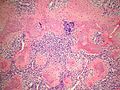
- "White clouds in a blue sky".
- Malignant tumor with a characteristic biphasic pattern
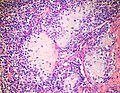
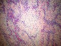
- Cellular poorly differentiated small round blue cells
- Islands of well-differentiated hyaline cartilage
- Progressive maturation of cartilage towards the center
- Central calcification or bone formation
IHC
- SOX9 (positive in small cells and chondrocytes) [3]
- S100 (positive in chondrocytes not in small cells)
- Osteocalcin (negative in small cells)
Molecular
t(8;8)(q21.1;q13.3) HEY1-NCOA2 [4]
Photos
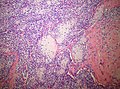
Islands of cartilage in a background of small blue cells.(SKB)
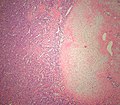
Islands of cartilage in a background of small blue cells.(SKB)
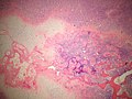
Islands of cartilage in a background of small blue cells.(SKB)
Islands of cartilage in a background of small blue cells.(SKB)
Islands of cartilage in a background of small blue cells.(SKB)
Islands of cartilage in a background of small blue cells.(SKB)

Small blue cells predominate.(SKB)


See also
Image:
References
- ↑ 1.0 1.1 Dowling EA (June 1964). "Mesenchymal chondrosarcoma". J Bone Joint Surg Am 46: 747–54. PMID 14161087. http://www.ejbjs.org/cgi/reprint/46/4/747.pdf.
- ↑ Fanburg-Smith, JC.; Auerbach, A.; Marwaha, JS.; Wang, Z.; Rushing, EJ. (May 2010). "Reappraisal of mesenchymal chondrosarcoma: novel morphologic observations of the hyaline cartilage and endochondral ossification and beta-catenin, Sox9, and osteocalcin immunostaining of 22 cases.". Hum Pathol 41 (5): 653-62. doi:10.1016/j.humpath.2009.11.006. PMID 20138330.
- ↑ Pang, ZG.; He, XZ.; Wu, LY.; Wei, W.; Liu, XY.; Liao, DY.; Li, FY.; Zhang, XL. (Jun 2011). "[Clinicopathologic and immunohistochemical study of 23 cases of mesenchymal chondrosarcoma].". Zhonghua Bing Li Xue Za Zhi 40 (6): 368-72. PMID 21914343.
- ↑ Panagopoulos, I.; Gorunova, L.; Bjerkehagen, B.; Boye, K.; Heim, S. (Jul 2014). "Chromosome aberrations and HEY1-NCOA2 fusion gene in a mesenchymal chondrosarcoma.". Oncol Rep 32 (1): 40-4. doi:10.3892/or.2014.3180. PMID 24839999.