Difference between revisions of "Malakoplakia"
Jump to navigation
Jump to search
m (→Microscopic: +image) |
(→Microscopic: more) |
||
| Line 12: | Line 12: | ||
*Occasional multinucleated giant cell. | *Occasional multinucleated giant cell. | ||
*Lymphocytes. | *Lymphocytes. | ||
DDx: | |||
*Xanthogranulomatous process. | |||
**If you cannot find the Michaelis-Gutmann bodies... it is a xanthogranulomatous process, e.g. [[xanthogranulomatous pyelonephritis]], xanthogranulomatous cystitis. | |||
Images: | Images: | ||
Revision as of 13:26, 17 August 2011
Malakoplakia is a thingy that typically arises in the bladder.
Gross
- Yellow mass.[1]
- May mimic renal cell carcinoma.
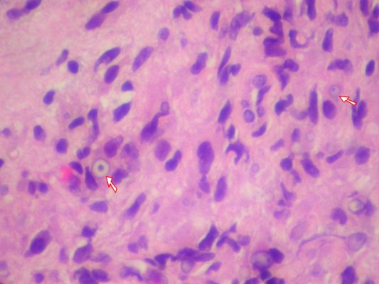
Microscopic
Features:[2]
- Basophilic calcified lysosomes (Michaelis-Gutmann bodies) -- key feature.
- May be inside or outside of macrophages - often size of RBC or larger.
- Large foamy macrophages with granular cytoplasm.
- Occasional multinucleated giant cell.
- Lymphocytes.
DDx:
- Xanthogranulomatous process.
- If you cannot find the Michaelis-Gutmann bodies... it is a xanthogranulomatous process, e.g. xanthogranulomatous pyelonephritis, xanthogranulomatous cystitis.
Images:
- Michaelis-Gutmann bodies - high mag. cropped (WC).
- Michaelis-Gutmann bodies - high mag. (WC).
- Malakoplakia (pathconsultddx.com)
- Malakoplakia (granuloma.homstead.com).
- Michaelis-Gutmann bodies (gfmer.ch).
See also
References
- ↑ URL: http://www.pathconsultddx.com/pathCon/diagnosis?pii=S1559-8675(06)70719-7. Accessed on: 9 September 2010.
- ↑ Cotran, Ramzi S.; Kumar, Vinay; Fausto, Nelson; Nelso Fausto; Robbins, Stanley L.; Abbas, Abul K. (2005). Robbins and Cotran pathologic basis of disease (7th ed.). St. Louis, Mo: Elsevier Saunders. pp. 1027. ISBN 0-7216-0187-1.