Difference between revisions of "Malakoplakia"
Jump to navigation
Jump to search
m (more) |
m (another image) |
||
| (12 intermediate revisions by one other user not shown) | |||
| Line 1: | Line 1: | ||
'''Malakoplakia''' is a thingy that typically arises in the bladder. | {{ Infobox diagnosis | ||
| Name = {{PAGENAME}} | |||
| Image = Michaelis-Gutmann_bodies_-_very_high_mag_-_cropped.jpg | |||
| Width = | |||
| Caption = Malakoplakia with numerous Michaelis-Gutmann bodies. [[H&E stain]]. | |||
| Micro = basophilic calcified bodies approximately the size of a RBC (Michaelis-Gutmann body) - inside or outside of macrophages, large foamy macrophages with granular cytoplasm +/- multinucleation, lymphocytes | |||
| Subtypes = | |||
| LMDDx = Xanthogranulomatous process (e.g. [[xanthogranulomatous pyelonephritis]], xanthogranulomatous cystitis), [[Granuloma|granulomatous inflammation]], [[papillary renal cell carcinoma]], infarction (prostate), [[granular cell tumour]] | |||
| Stains = | |||
| IHC = | |||
| EM = | |||
| Molecular = | |||
| IF = | |||
| Gross = yellow mass | |||
| Grossing = | |||
| Site = [[urinary bladder]] and elsewhere | |||
| Assdx = | |||
| Syndromes = | |||
| Clinicalhx = | |||
| Signs = | |||
| Symptoms = | |||
| Prevalence = uncommon | |||
| Bloodwork = | |||
| Rads = | |||
| Endoscopy = yellow lesion | |||
| Prognosis = benign | |||
| Other = | |||
| ClinDDx = [[renal cell carcinoma]], other tumours | |||
}} | |||
'''Malakoplakia''' is a thingy that typically arises in the [[urinary bladder]]. | |||
==General== | ==General== | ||
*Classically in the [[urinary bladder]]. | *Classically in the [[urinary bladder]]. | ||
**May be | **May be outside of urinary tract.<ref name=pmid17284117>{{Cite journal | last1 = Yousef | first1 = GM. | last2 = Naghibi | first2 = B. | last3 = Hamodat | first3 = MM. | title = Malakoplakia outside the urinary tract. | journal = Arch Pathol Lab Med | volume = 131 | issue = 2 | pages = 297-300 | month = Feb | year = 2007 | doi = 10.1043/1543-2165(2007)131[297:MOTUT]2.0.CO;2 | PMID = 17284117 }}</ref> | ||
==Gross== | ==Gross== | ||
*Yellow mass.<ref>URL: [http://www.pathconsultddx.com/pathCon/diagnosis?pii=S1559-8675(06)70719-7 http://www.pathconsultddx.com/pathCon/diagnosis?pii=S1559-8675(06)70719-7]. Accessed on: 9 September 2010.</ref> | *Yellow mass.<ref>URL: [http://www.pathconsultddx.com/pathCon/diagnosis?pii=S1559-8675(06)70719-7 http://www.pathconsultddx.com/pathCon/diagnosis?pii=S1559-8675(06)70719-7]. Accessed on: 9 September 2010.</ref> | ||
**May mimic renal cell carcinoma. | **May mimic [[renal cell carcinoma]]. | ||
==Microscopic== | ==Microscopic== | ||
| Line 20: | Line 49: | ||
*Xanthogranulomatous process. | *Xanthogranulomatous process. | ||
**If you cannot find the Michaelis-Gutmann bodies... it is a xanthogranulomatous process, e.g. [[xanthogranulomatous pyelonephritis]], xanthogranulomatous cystitis. | **If you cannot find the Michaelis-Gutmann bodies... it is a xanthogranulomatous process, e.g. [[xanthogranulomatous pyelonephritis]], xanthogranulomatous cystitis. | ||
*Granulomatous inflammation. | *[[Granuloma|Granulomatous inflammation]]. | ||
* | *[[Papillary renal cell carcinoma]]. | ||
*Infarction (prostate). | |||
*[[Granular cell tumour]].<ref name=pmid19239951/> | |||
Images | ===Images=== | ||
====Case==== | |||
<gallery> | |||
Image:Michaelis-Gutmann_bodies_-_very_high_mag_-_cropped.jpg | Michaelis-Gutmann bodies - high mag. cropped (WC) | |||
Image:Michaelis-Gutmann_bodies_-_very_high_mag.jpg | Michaelis-Gutmann bodies - high mag. (WC) | |||
Image:malakoplakia bladder.jpg | Michaelis-Gutmann bodies - high mag. (WC) | |||
</gallery> | |||
====www==== | |||
*[http://granuloma.homestead.com/malakoplakia_bladder_s92-4486e.jpg Malakoplakia (granuloma.homstead.com)]. | *[http://granuloma.homestead.com/malakoplakia_bladder_s92-4486e.jpg Malakoplakia (granuloma.homstead.com)]. | ||
*[http://www.gfmer.ch/selected_images_v2/detail_list.php?cat1=13&cat3=342&stype=d Michaelis-Gutmann bodies (gfmer.ch)]. | *[http://www.gfmer.ch/selected_images_v2/detail_list.php?cat1=13&cat3=342&stype=d Michaelis-Gutmann bodies (gfmer.ch)]. | ||
==Stains== | |||
*[[PAS-D stain]] +ve.<ref name=pmid17284117/> | |||
*[[Von Kossa stain]] +ve.<ref name=pmid19239951>{{Cite journal | last1 = Diapera | first1 = MJ. | last2 = Lozon | first2 = CL. | last3 = Thompson | first3 = LD. | title = Malacoplakia of the tongue: a case report and clinicopathologic review of 6 cases. | journal = Am J Otolaryngol | volume = 30 | issue = 2 | pages = 101-5 | month = | year = | doi = 10.1016/j.amjoto.2008.02.014 | PMID = 19239951 }}</ref> | |||
==See also== | ==See also== | ||
| Line 39: | Line 79: | ||
[[Category: Genitourinary pathology]] | [[Category: Genitourinary pathology]] | ||
[[Category:Diagnosis]] | |||
[[Category:Urothelium]] | |||
Latest revision as of 15:51, 30 September 2018
| Malakoplakia | |
|---|---|
| Diagnosis in short | |
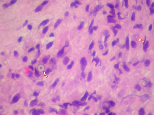
 Malakoplakia with numerous Michaelis-Gutmann bodies. H&E stain. | |
|
| |
| LM | basophilic calcified bodies approximately the size of a RBC (Michaelis-Gutmann body) - inside or outside of macrophages, large foamy macrophages with granular cytoplasm +/- multinucleation, lymphocytes |
| LM DDx | Xanthogranulomatous process (e.g. xanthogranulomatous pyelonephritis, xanthogranulomatous cystitis), granulomatous inflammation, papillary renal cell carcinoma, infarction (prostate), granular cell tumour |
| Gross | yellow mass |
| Site | urinary bladder and elsewhere |
|
| |
| Prevalence | uncommon |
| Endoscopy | yellow lesion |
| Prognosis | benign |
| Clin. DDx | renal cell carcinoma, other tumours |
Malakoplakia is a thingy that typically arises in the urinary bladder.
General
- Classically in the urinary bladder.
- May be outside of urinary tract.[1]
Gross
- Yellow mass.[2]
- May mimic renal cell carcinoma.
Microscopic
Features:[3]
- Basophilic calcified lysosomes (Michaelis-Gutmann bodies) -- key feature.
- May be inside or outside of macrophages - often size of RBC or larger.
- Large foamy macrophages with granular cytoplasm.
- Occasional multinucleated giant cell.
- Lymphocytes.
DDx:
- Xanthogranulomatous process.
- If you cannot find the Michaelis-Gutmann bodies... it is a xanthogranulomatous process, e.g. xanthogranulomatous pyelonephritis, xanthogranulomatous cystitis.
- Granulomatous inflammation.
- Papillary renal cell carcinoma.
- Infarction (prostate).
- Granular cell tumour.[4]
Images
Case
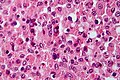
Michaelis-Gutmann bodies - high mag. cropped (WC)
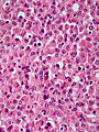
Michaelis-Gutmann bodies - high mag. (WC)
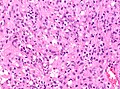
Michaelis-Gutmann bodies - high mag. (WC)


www
Stains
- PAS-D stain +ve.[1]
- Von Kossa stain +ve.[4]
See also
References
- ↑ 1.0 1.1 Yousef, GM.; Naghibi, B.; Hamodat, MM. (Feb 2007). "Malakoplakia outside the urinary tract.". Arch Pathol Lab Med 131 (2): 297-300. doi:10.1043/1543-2165(2007)131[297:MOTUT]2.0.CO;2. PMID 17284117.
- ↑ URL: http://www.pathconsultddx.com/pathCon/diagnosis?pii=S1559-8675(06)70719-7. Accessed on: 9 September 2010.
- ↑ Cotran, Ramzi S.; Kumar, Vinay; Fausto, Nelson; Nelso Fausto; Robbins, Stanley L.; Abbas, Abul K. (2005). Robbins and Cotran pathologic basis of disease (7th ed.). St. Louis, Mo: Elsevier Saunders. pp. 1027. ISBN 0-7216-0187-1.
- ↑ 4.0 4.1 Diapera, MJ.; Lozon, CL.; Thompson, LD.. "Malacoplakia of the tongue: a case report and clinicopathologic review of 6 cases.". Am J Otolaryngol 30 (2): 101-5. doi:10.1016/j.amjoto.2008.02.014. PMID 19239951.