Difference between revisions of "Lung metastasis"
Jump to navigation
Jump to search
(→Microscopic: +ddx) |
|||
| Line 7: | Line 7: | ||
| Micro = | | Micro = | ||
| Subtypes = | | Subtypes = | ||
| LMDDx = primary lung cancer | | LMDDx = primary lung cancer ([[adenocarcinoma of the lung]], [[squamous cell carcinoma of the lung]], [[small cell carcinoma of the lung]]), [[pulmonary meningothelial-like nodule]], [[carcinoid tumourlet]], [[carcinoid lung tumour]] | ||
| Stains = | | Stains = | ||
| IHC = TTF-1 (-ve useful if non-squamous), CK20 (+ve suggestive colorectal carcinoma), CK7 (-ve useful if non-squamous), GATA3 (+ve suggestive UCC) | | IHC = TTF-1 (-ve useful if non-squamous), CK20 (+ve suggestive colorectal carcinoma), CK7 (-ve useful if non-squamous), GATA3 (+ve suggestive UCC) | ||
| Line 27: | Line 27: | ||
| Prognosis = usually poor | | Prognosis = usually poor | ||
| Other = | | Other = | ||
| ClinDDx = lung primary, abscess | | ClinDDx = lung primary, abscess, multiple benign lung tumours as may be seen in [[DIPNECH]] | ||
| Tx = | | Tx = dependent on primary site, occasionally surgical | ||
}} | }} | ||
'''Lung metastasis''', also '''pulmonary metastasis''' and '''metastatic lung disease''', is relatively common and generally carries a poor prognosis. | '''Lung metastasis''', also '''pulmonary metastasis''' and '''metastatic lung disease''', is relatively common and generally carries a poor prognosis. | ||
| Line 56: | Line 56: | ||
DDx: | DDx: | ||
*Primary lung tumour, e.g. [[lung adenocarcinoma]], [[lung squamous cell carcinoma]], [[small cell carcinoma of the lung]]. | |||
*[[Pulmonary meningothelial-like nodule]].<ref name=pmid23109985>{{Cite journal | last1 = Kfoury | first1 = H. | last2 = Arafah | first2 = MA. | last3 = Arafah | first3 = MM. | last4 = Alnassar | first4 = S. | last5 = Hajjar | first5 = W. | title = Mimicry of Minute Pulmonary Meningothelial-like Nodules to Metastatic Deposits in a Patient with Infiltrating Lobular Carcinoma: A Case Report and Review of the Literature. | journal = Korean J Pathol | volume = 46 | issue = 1 | pages = 87-91 | month = Feb | year = 2012 | doi = 10.4132/KoreanJPathol.2012.46.1.87 | PMID = 23109985 }}</ref> | *[[Pulmonary meningothelial-like nodule]].<ref name=pmid23109985>{{Cite journal | last1 = Kfoury | first1 = H. | last2 = Arafah | first2 = MA. | last3 = Arafah | first3 = MM. | last4 = Alnassar | first4 = S. | last5 = Hajjar | first5 = W. | title = Mimicry of Minute Pulmonary Meningothelial-like Nodules to Metastatic Deposits in a Patient with Infiltrating Lobular Carcinoma: A Case Report and Review of the Literature. | journal = Korean J Pathol | volume = 46 | issue = 1 | pages = 87-91 | month = Feb | year = 2012 | doi = 10.4132/KoreanJPathol.2012.46.1.87 | PMID = 23109985 }}</ref> | ||
*[[Carcinoid tumourlet]]. | *[[Carcinoid tumourlet]]. | ||
Revision as of 15:31, 28 September 2015
| Lung metastasis | |
|---|---|
| Diagnosis in short | |
 Lung metastasis (Ewing sarcoma). H&E stain. | |
| LM DDx | primary lung cancer (adenocarcinoma of the lung, squamous cell carcinoma of the lung, small cell carcinoma of the lung), pulmonary meningothelial-like nodule, carcinoid tumourlet, carcinoid lung tumour |
| IHC | TTF-1 (-ve useful if non-squamous), CK20 (+ve suggestive colorectal carcinoma), CK7 (-ve useful if non-squamous), GATA3 (+ve suggestive UCC) |
| Gross | lung nodules - typically multiple and peripheral |
| Site | lung |
|
| |
| Clinical history | +/-hx of cancer |
| Prevalence | relatively common |
| Radiology | peripheral lung lesions, typically multiple |
| Prognosis | usually poor |
| Clin. DDx | lung primary, abscess, multiple benign lung tumours as may be seen in DIPNECH |
| Treatment | dependent on primary site, occasionally surgical |
Lung metastasis, also pulmonary metastasis and metastatic lung disease, is relatively common and generally carries a poor prognosis.
General
- Relatively common.
Gross
- Typically peripheral, multiple, well-circumscribed & white/tan masses.
- May be diffuse without an obvious mass +/- septal thickening.
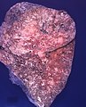
Prostate carcinoma. (WC/Rosen)
.jpg)
Microscopic
Features:
- Variable - dependent on site of origin.
- Colorectal adenocarcinoma - usually distinctive morphologically:
- Typically gland forming.
- Ellipsoid/elongated pseudostratified nuclei with moderate nuclear atypia.
- +/-Dirty necrosis.
- Typically gland forming.
- Others:
- Urothelial carcinoma - may mimic squamous cell carcinoma of the lung.
- Upper GI adenocarcinoma (e.g. gastric adenocarcinoma) - may mimic lung adenocarcinoma.
- Breast carcinoma - esp. ductal carcinoma of the breast - may mimic lung adenocarcinoma.
DDx:
- Primary lung tumour, e.g. lung adenocarcinoma, lung squamous cell carcinoma, small cell carcinoma of the lung.
- Pulmonary meningothelial-like nodule.[1]
- Carcinoid tumourlet.
- Carcinoid lung tumour.
Images
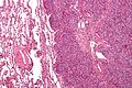
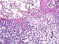
Lung metastasis (ES) - intermed. mag. (WC/Nephron)
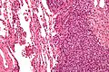
Lung metastasis (ES) - high mag. (WC/Nephron)
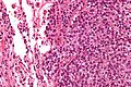
Lung metastasis (ES) - very high mag. (WC/Nephron)


Prostate carcinoma. (WC/Rosen)
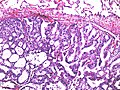
Prostate carcinoma. (WC/Rosen)
.jpg)
.jpg)
IHC
- TTF-1 -ve/+ve.
- Negative suggestive of metastasis... unless it is squamous carcinoma.
- CK20 +ve/-ve.
- Positive in colorectal carcinoma - very useful.
- Negative in lung primaries.
- GATA3 +ve/-ve.
- Usu. +ve in urothelial carcinoma.
- Negative in lung primaries.[2]
- CK7 -ve/+ve.
- Positive in lung adenocarcinoma and small carcinoma of the lung.
- Positive in a number of other tumours - breast, upper GI tract, thyroid, mesothelioma, salivary gland.
- Negative in poorly differentiated carcinoma of the lung and squamous carcinoma of the lung.
See also
References
- ↑ Kfoury, H.; Arafah, MA.; Arafah, MM.; Alnassar, S.; Hajjar, W. (Feb 2012). "Mimicry of Minute Pulmonary Meningothelial-like Nodules to Metastatic Deposits in a Patient with Infiltrating Lobular Carcinoma: A Case Report and Review of the Literature.". Korean J Pathol 46 (1): 87-91. doi:10.4132/KoreanJPathol.2012.46.1.87. PMID 23109985.
- ↑ Chang, A.; Amin, A.; Gabrielson, E.; Illei, P.; Roden, RB.; Sharma, R.; Epstein, JI. (Oct 2012). "Utility of GATA3 immunohistochemistry in differentiating urothelial carcinoma from prostate adenocarcinoma and squamous cell carcinomas of the uterine cervix, anus, and lung.". Am J Surg Pathol 36 (10): 1472-6. doi:10.1097/PAS.0b013e318260cde7. PMID 22982890.