Difference between revisions of "Inflammatory myofibroblastic tumour"
Jump to navigation
Jump to search
| Line 1: | Line 1: | ||
{{ Infobox diagnosis | |||
| Name = {{PAGENAME}} | |||
| Image = Inflammatory_myofibroblastic_tumour_-_very_high_mag.jpg | |||
| Width = | |||
| Caption = Inflammatory myofibroblastic tumour. [[H&E stain]]. | |||
| Synonyms = | |||
| Micro = inflammation ([[plasma cells]] - predominant, lymphocytes, eosinophils), [[spindle cells]] without atypia +/-fascicular architecture, +/-mitoses (none atypical), +/-[[necrosis]], +/-hemorrhage, +/-calcification | |||
| Subtypes = | |||
| LMDDx = [[calcifying fibrous pseudotumour]], [[inflammatory fibroid tumour]], [[nodular fasciitis]] | |||
| Stains = | |||
| IHC = | |||
| EM = | |||
| Molecular = | |||
| IF = | |||
| Gross = | |||
| Grossing = | |||
| Site = [[soft tissue lesions|soft tissue]] - see ''[[fibroblastic/myofibroblastic tumours]] | |||
| Assdx = | |||
| Syndromes = | |||
| Clinicalhx = | |||
| Signs = | |||
| Symptoms = | |||
| Prevalence = uncommon | |||
| Bloodwork = | |||
| Rads = | |||
| Endoscopy = | |||
| Prognosis = benign | |||
| Other = | |||
| ClinDDx = other soft tissue lesions | |||
| Tx = | |||
}} | |||
'''Inflammatory myofibroblastic tumour''' is an uncommon [[soft tissue lesion]]. | '''Inflammatory myofibroblastic tumour''' is an uncommon [[soft tissue lesion]]. | ||
| Line 15: | Line 46: | ||
**Eosinophils. | **Eosinophils. | ||
*Spindle cells without atypia. | *Spindle cells without atypia. | ||
*+/- | *+/-Fascicular architecture. | ||
*Mitoses -- though none atypical. | *Mitoses -- though none atypical. | ||
*+/-Necrosis. | *+/-Necrosis. | ||
Revision as of 04:42, 13 August 2014
| Inflammatory myofibroblastic tumour | |
|---|---|
| Diagnosis in short | |
 Inflammatory myofibroblastic tumour. H&E stain. | |
|
| |
| LM | inflammation (plasma cells - predominant, lymphocytes, eosinophils), spindle cells without atypia +/-fascicular architecture, +/-mitoses (none atypical), +/-necrosis, +/-hemorrhage, +/-calcification |
| LM DDx | calcifying fibrous pseudotumour, inflammatory fibroid tumour, nodular fasciitis |
| Site | soft tissue - see fibroblastic/myofibroblastic tumours |
|
| |
| Prevalence | uncommon |
| Prognosis | benign |
| Clin. DDx | other soft tissue lesions |
Inflammatory myofibroblastic tumour is an uncommon soft tissue lesion.
It is also known as inflammatory pseudotumour, and inflammatory fibrosarcoma[1] and plasma cell granuloma.[2][3]
General
- Mostly benign.
- Children & young adults.
- Classically located in mesentery of ileocolic region or small bowel.[1]
Microscopic
Features:[1]
- Inflammation:
- Plasma cells - predominant - key feature.[4]
- Lymphocytes.
- Eosinophils.
- Spindle cells without atypia.
- +/-Fascicular architecture.
- Mitoses -- though none atypical.
- +/-Necrosis.
- +/-Hemorrhage.
- Calcifications.
DDx:
- Calcifying fibrous pseudotumour (has psammomatous calcifications).
- Inflammatory fibroid tumour.
- Nodular fasciitis.
Notes:
- Some consider this a wastebasket diagnosis... for benign appearing spindle cell lesions.[5]
Images
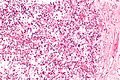
IMT - high mag. (WC)
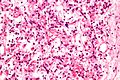
IMT - very high mag. (WC)

IHC
Features - dependent on site:
- SMA +ve.[6]
- Vimentin +ve.
Variable staining with:
Negative:[6]
- S100, CD117, CD68.
Others:
- ALK-1 +ve. ???
Molecular
- ALK rearrangements.[4]
See also
References
- ↑ 1.0 1.1 1.2 Humphrey, Peter A; Dehner, Louis P; Pfeifer, John D (2008). The Washington Manual of Surgical Pathology (1st ed.). Lippincott Williams & Wilkins. pp. 610. ISBN 978-0781765275.
- ↑ URL: http://www.uptodate.com/contents/inflammatory-myofibroblastic-tumor-plasma-cell-granuloma-of-the-lung. Accessed on: 27 November 2011.
- ↑ Manohar, B.; Bhuvaneshwari, S. (Jan 2011). "Plasma cell granuloma of gingiva.". J Indian Soc Periodontol 15 (1): 64-6. doi:10.4103/0972-124X.82275. PMID 21772725.
- ↑ 4.0 4.1 Saab, ST.; Hornick, JL.; Fletcher, CD.; Olson, SJ.; Coffin, CM. (Apr 2011). "IgG4 plasma cells in inflammatory myofibroblastic tumor: inflammatory marker or pathogenic link?". Mod Pathol 24 (4): 606-12. doi:10.1038/modpathol.2010.226. PMID 21297584.
- ↑ URL: http://www.pathconsultddx.com/pathCon/diagnosis?pii=S1559-8675%2806%2970283-2. Accessed on: 10 May 2011.
- ↑ 6.0 6.1 6.2 Shi, H.; Li, Y.; Wei, L.; Sun, L. (Apr 2010). "Primary colorectal inflammatory myofibroblastic tumour: a clinicopathological and immunohistochemical study of seven cases.". Pathology 42 (3): 235-41. doi:10.3109/00313021003631312. PMID 20350216.
Cite error: Invalid
<ref>tag; name "pmid20350216" defined multiple times with different content - ↑ Miyamoto, H.; Montgomery, EA.; Epstein, JI. (Apr 2010). "Paratesticular fibrous pseudotumor: a morphologic and immunohistochemical study of 13 cases.". Am J Surg Pathol 34 (4): 569-74. doi:10.1097/PAS.0b013e3181d438cb. PMID 20216379.