Difference between revisions of "Giant cell tumour of bone"
Jump to navigation
Jump to search
(fix redirect) |
(more) |
||
| (7 intermediate revisions by the same user not shown) | |||
| Line 1: | Line 1: | ||
{{ Infobox diagnosis | |||
| Name = {{PAGENAME}} | |||
| Image = Giant_cell_tumour_of_bone_-_high_mag.jpg | |||
| Width = | |||
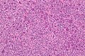
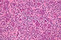
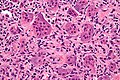
| Caption = Giant cell tumour of bone. [[H&E stain]]. | |||
| Micro = giant cells (usu. with >10 in the plane of section), mononuclear cells and small multinucleated cells with nuclei similar to those in the giant cells | |||
| Subtypes = | |||
| LMDDx = other [[giant cell lesions]] | |||
| Stains = | |||
| IHC = p63 +ve (patchy) | |||
| EM = | |||
| Molecular = | |||
| IF = | |||
| Gross = | |||
| Grossing = | |||
| Site = [[bone]], epiphysis usually | |||
| Assdx = | |||
| Syndromes = | |||
| Clinicalhx = usu. young adults (20-45 years old) | |||
| Signs = +/-immobility | |||
| Symptoms = +/-pain | |||
| Prevalence = uncommon ~ 5% of primary bone tumours | |||
| Bloodwork = | |||
| Rads = | |||
| Endoscopy = | |||
| Prognosis = | |||
| Other = | |||
| ClinDDx = | |||
}} | |||
'''Giant cell tumour of bone''' is an uncommon [[bone]] tumour. | |||
==General== | |||
Features:<ref name=Ref_WMSP648>{{Ref WMSP|648}}</ref> | |||
*Approximately 5% of primary [[bone tumour]]s. | |||
*Typical age: 20-45 years. | |||
===Clinical=== | |||
*Location: growth plate of long bones.<ref name=pmid11501745>{{Cite journal | last1 = Wülling | first1 = M. | last2 = Engels | first2 = C. | last3 = Jesse | first3 = N. | last4 = Werner | first4 = M. | last5 = Delling | first5 = G. | last6 = Kaiser | first6 = E. | title = The nature of giant cell tumor of bone. | journal = J Cancer Res Clin Oncol | volume = 127 | issue = 8 | pages = 467-74 | month = Aug | year = 2001 | doi = | PMID = 11501745 }}</ref> | |||
**May present with joint pain, immobility. | |||
Note: | |||
*Several types of [[giant cell lesions|giant cell tumours]] exist. | |||
==Microscopic== | |||
Features:<ref name=Ref_Klatt420>{{Ref Klatt|420}}</ref> | |||
*Giant cells with a large number of nuclei (usu. >10 in the plane of section). | |||
**Usu. have [[prominent nucleoli]]. | |||
*Mononuclear cells and small multinucleated cells with nuclei similar to those in the giant cells - '''key feature'''. | |||
*+/-Hemosiderin deposition - not common.<ref name=pmid1939753>{{Cite journal | last1 = Aoki | first1 = J. | last2 = Moriya | first2 = K. | last3 = Yamashita | first3 = K. | last4 = Fujioka | first4 = F. | last5 = Ishii | first5 = K. | last6 = Karakida | first6 = O. | last7 = Imai | first7 = S. | last8 = Sakai | first8 = F. | last9 = Imai | first9 = Y. | title = Giant cell tumors of bone containing large amounts of hemosiderin: MR-pathologic correlation. | journal = J Comput Assist Tomogr | volume = 15 | issue = 6 | pages = 1024-7 | month = | year = | doi = | PMID = 1939753 }}</ref><ref name=pmid18554912>{{Cite journal | last1 = Matsushige | first1 = T. | last2 = Nakaoka | first2 = M. | last3 = Yahara | first3 = K. | last4 = Kagawa | first4 = K. | last5 = Miura | first5 = H. | last6 = Ohnuma | first6 = H. | last7 = Kurisu | first7 = K. | title = Giant cell tumor of the temporal bone with intratumoral hemorrhage. | journal = J Clin Neurosci | volume = 15 | issue = 8 | pages = 923-7 | month = Aug | year = 2008 | doi = 10.1016/j.jocn.2007.03.013 | PMID = 18554912 }}</ref> | |||
Notes: | |||
*Giant cells typically present in abundance. | |||
DDx: | |||
*[[Giant cell lesions]]. | |||
**[[Aneurysmal bone cyst]] - typically has spindle cells around the giant cells. | |||
===Images=== | |||
<gallery> | |||
Image:Giant_cell_tumour_of_bone_-_low_mag.jpg | GCT of bone - low mag. (WC) | |||
Image:Giant cell tumour of bone - intermed mag.jpg | GCT of bone - intermed. mag. (WC) | |||
Image:Giant_cell_tumour_of_bone_-_high_mag.jpg | GCT of bone - high mag. (WC) | |||
Image:Giant cell tumour of bone - very high mag.jpg | GCT of bone - very high mag. (WC) | |||
</gallery> | |||
==IHC== | |||
*p63 +ve in scattered mononuclear cells.<ref name=pmid18311114>{{cite journal |author=Dickson BC, Li SQ, Wunder JS, ''et al.'' |title=Giant cell tumor of bone express p63 |journal=Mod. Pathol. |volume=21 |issue=4 |pages=369–75 |year=2008 |month=April |pmid=18311114 |doi=10.1038/modpathol.2008.29 |url=}}</ref> | |||
**This seems to be contradicted by another paper.<ref name=pmid20012988>{{cite journal |author=Alberghini M, Kliskey K, Krenacs T, ''et al.'' |title=Morphological and immunophenotypic features of primary and metastatic giant cell tumour of bone |journal=Virchows Arch. |volume=456 |issue=1 |pages=97–103 |year=2010 |month=January |pmid=20012988 |doi=10.1007/s00428-009-0863-2 |url=}}</ref> | |||
==See also== | |||
*[[Bone]]. | |||
*[[Chondro-osseous tumours]]. | |||
*[[Giant cells]]. | |||
*[[Giant cell lesions]]. | |||
==References== | |||
{{Reflist|2}} | |||
[[Category:Diagnosis]] | |||
[[Category:Chondro-osseous tumours]] | |||
Latest revision as of 07:42, 1 October 2013
| Giant cell tumour of bone | |
|---|---|
| Diagnosis in short | |
 Giant cell tumour of bone. H&E stain. | |
|
| |
| LM | giant cells (usu. with >10 in the plane of section), mononuclear cells and small multinucleated cells with nuclei similar to those in the giant cells |
| LM DDx | other giant cell lesions |
| IHC | p63 +ve (patchy) |
| Site | bone, epiphysis usually |
|
| |
| Clinical history | usu. young adults (20-45 years old) |
| Signs | +/-immobility |
| Symptoms | +/-pain |
| Prevalence | uncommon ~ 5% of primary bone tumours |
Giant cell tumour of bone is an uncommon bone tumour.
General
Features:[1]
- Approximately 5% of primary bone tumours.
- Typical age: 20-45 years.
Clinical
- Location: growth plate of long bones.[2]
- May present with joint pain, immobility.
Note:
- Several types of giant cell tumours exist.
Microscopic
Features:[3]
- Giant cells with a large number of nuclei (usu. >10 in the plane of section).
- Usu. have prominent nucleoli.
- Mononuclear cells and small multinucleated cells with nuclei similar to those in the giant cells - key feature.
- +/-Hemosiderin deposition - not common.[4][5]
Notes:
- Giant cells typically present in abundance.
DDx:
- Giant cell lesions.
- Aneurysmal bone cyst - typically has spindle cells around the giant cells.
Images
GCT of bone - low mag. (WC)
GCT of bone - intermed. mag. (WC)
GCT of bone - high mag. (WC)
GCT of bone - very high mag. (WC)


IHC
See also
References
- ↑ Humphrey, Peter A; Dehner, Louis P; Pfeifer, John D (2008). The Washington Manual of Surgical Pathology (1st ed.). Lippincott Williams & Wilkins. pp. 648. ISBN 978-0781765275.
- ↑ Wülling, M.; Engels, C.; Jesse, N.; Werner, M.; Delling, G.; Kaiser, E. (Aug 2001). "The nature of giant cell tumor of bone.". J Cancer Res Clin Oncol 127 (8): 467-74. PMID 11501745.
- ↑ Klatt, Edward C. (2006). Robbins and Cotran Atlas of Pathology (1st ed.). Saunders. pp. 420. ISBN 978-1416002741.
- ↑ Aoki, J.; Moriya, K.; Yamashita, K.; Fujioka, F.; Ishii, K.; Karakida, O.; Imai, S.; Sakai, F. et al. "Giant cell tumors of bone containing large amounts of hemosiderin: MR-pathologic correlation.". J Comput Assist Tomogr 15 (6): 1024-7. PMID 1939753.
- ↑ Matsushige, T.; Nakaoka, M.; Yahara, K.; Kagawa, K.; Miura, H.; Ohnuma, H.; Kurisu, K. (Aug 2008). "Giant cell tumor of the temporal bone with intratumoral hemorrhage.". J Clin Neurosci 15 (8): 923-7. doi:10.1016/j.jocn.2007.03.013. PMID 18554912.
- ↑ Dickson BC, Li SQ, Wunder JS, et al. (April 2008). "Giant cell tumor of bone express p63". Mod. Pathol. 21 (4): 369–75. doi:10.1038/modpathol.2008.29. PMID 18311114.
- ↑ Alberghini M, Kliskey K, Krenacs T, et al. (January 2010). "Morphological and immunophenotypic features of primary and metastatic giant cell tumour of bone". Virchows Arch. 456 (1): 97–103. doi:10.1007/s00428-009-0863-2. PMID 20012988.