Diversion colitis
Jump to navigation
Jump to search
The printable version is no longer supported and may have rendering errors. Please update your browser bookmarks and please use the default browser print function instead.
| Diversion colitis | |
|---|---|
| Diagnosis in short | |
 Diversion proctitis. H&E stain. | |
|
| |
| LM | follicular lymphoid hyperplasia (abundant lymphoid nodules, plasma cells), +/-changes of an active colitis (cryptitis, crypt abscesses) - uncommon |
| LM DDx | inflammatory bowel disease - no stoma, ischemic colitis, infectious colitis, lymphoma |
| Site | colon, rectum |
|
| |
| Clinical history | previous diversion - history essential |
| Prevalence | uncommon |
| Prognosis | usu. resolves with re-anastomosis |
| Clin. DDx | other causes of colitis |
Diversion colitis is colitis due to a diversion, i.e. a stoma. Diversion proctitis redirects here.
General
- Segment of de-functioned bowel due to surgical diversion, i.e. stoma (ileostomy or colostomy).
- Diagnosis dependent on history - key point.
Gross
Features:[1]
- Ulceration - classic.
- Surgical changes, e.g. fibrotic-appearing thickened wall.
- May not be apparent.
Microscopic
Features:[1]
- Follicular lymphoid hyperplasia - key feature.[2]
- Abundant lymphoid nodules.
- Plasma cells and lymphocytes.
- +/-Changes of an active colitis - uncommon:[3]
Notes:
- May show IBD-like changes.[4]
- IBD should not be diagnosed on a diverted segment of bowel.
DDx:[5]
- Inflammatory bowel disease - no stoma.
- Ischemic colitis.
- Infectious colitis.
Images
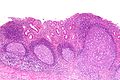
Diversion proctitis - low mag. (WC/Nephron)
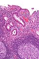
Diversion proctitis - high mag. (WC/Nephron)

Sign out
SIGMOID COLON, BIOPSIES:
- MILD ACTIVE COLITIS WITH LAMINA PROPRIA FIBROSIS, SEE COMMENT.
- NEGATIVE FOR DYSPLASIA.

COMMENT:
No granulomas are identified.  Follicular lymphoid hyperplasia is not identified;
however, there is no definite submucosa present.

Diverted segments of bowel can have inflammatory bowel disease-like changes.

In the context of a diverted segment of bowel, the findings are compatible with
a diversion colitis.
RECTUM, BIOPSY:
- CHRONIC ACTIVE PROCTITIS WITH FOCAL ULCERATION, CRYPTITIS AND CRYPT ABSCESSES.
- GRANULATION TISSUE.
- NEGATIVE FOR DYSPLASIA.

COMMENT:
No lymphoid hyperplasia is present. A small lymphoid aggregate is present.
Architectural distortion is present.

In the context of a diverted segment of bowel, the findings are compatible with
a diversion colitis.
See also
References
- ↑ 1.0 1.1 Edwards, CM.; George, B.; Warren, B. (Jan 1999). "Diversion colitis--new light through old windows.". Histopathology 34 (1): 1-5. PMID 9934577.
- ↑ Yeong, ML.; Bethwaite, PB.; Prasad, J.; Isbister, WH. (Jul 1991). "Lymphoid follicular hyperplasia--a distinctive feature of diversion colitis.". Histopathology 19 (1): 55-61. PMID 1916687.
- ↑ Ma, CK.; Gottlieb, C.; Haas, PA. (Apr 1990). "Diversion colitis: a clinicopathologic study of 21 cases.". Hum Pathol 21 (4): 429-36. PMID 2318485.
- ↑ Yantiss, RK.; Odze, RD. (Jan 2006). "Diagnostic difficulties in inflammatory bowel disease pathology.". Histopathology 48 (2): 116-32. doi:10.1111/j.1365-2559.2005.02248.x. PMID 16405661.
- ↑ Thorsen, AJ. (Feb 2007). "Noninfectious colitides: collagenous colitis, lymphocytic colitis, diversion colitis, and chemically induced colitis.". Clin Colon Rectal Surg 20 (1): 47-57. doi:10.1055/s-2007-970200. PMC 2780148. PMID 20011361. http://www.ncbi.nlm.nih.gov/pmc/articles/PMC2780148/.