Difference between revisions of "Diversion colitis"
Jump to navigation
Jump to search
(create redirect) |
|||
| (8 intermediate revisions by the same user not shown) | |||
| Line 1: | Line 1: | ||
{{ Infobox diagnosis | |||
| Name = {{PAGENAME}} | |||
| Image = Diversion_proctitis_-_low_mag.jpg | |||
| Width = | |||
| Caption = Diversion proctitis. [[H&E stain]]. | |||
| Micro = follicular lymphoid hyperplasia (abundant lymphoid nodules, plasma cells), +/-changes of an active colitis ([[cryptitis]], crypt abscesses) - uncommon | |||
| Subtypes = | |||
| LMDDx = [[inflammatory bowel disease]] - no stoma, [[ischemic colitis]], [[infectious colitis]], [[lymphoma]] | |||
| Stains = | |||
| IHC = | |||
| EM = | |||
| Molecular = | |||
| IF = | |||
| Gross = | |||
| Grossing = | |||
| Site = [[colon]], [[rectum]] | |||
| Assdx = | |||
| Syndromes = | |||
| Clinicalhx = previous diversion - history essential | |||
| Signs = | |||
| Symptoms = | |||
| Prevalence = uncommon | |||
| Bloodwork = | |||
| Rads = | |||
| Endoscopy = | |||
| Prognosis = usu. resolves with re-anastomosis | |||
| Other = | |||
| ClinDDx = other causes of [[colitis]] | |||
}} | |||
'''Diversion colitis''' is [[colitis]] due to a diversion, i.e. a [[stoma]]. '''Diversion proctitis''' redirects here. | |||
==General== | |||
*Segment of de-functioned bowel due to surgical diversion, i.e. stoma (ileostomy or [[colostomy]]). | |||
*[[Diagnosis]] dependent on history - '''key point'''. | |||
==Gross== | |||
Features:<ref name=pmid9934577/> | |||
*Ulceration - classic. | |||
*Surgical changes, e.g. fibrotic-appearing thickened wall. | |||
**May not be apparent. | |||
==Microscopic== | |||
Features:<ref name=pmid9934577>{{Cite journal | last1 = Edwards | first1 = CM. | last2 = George | first2 = B. | last3 = Warren | first3 = B. | title = Diversion colitis--new light through old windows. | journal = Histopathology | volume = 34 | issue = 1 | pages = 1-5 | month = Jan | year = 1999 | doi = | PMID = 9934577 }}</ref> | |||
*Follicular lymphoid hyperplasia - '''key feature'''.<ref name=pmid1916687>{{Cite journal | last1 = Yeong | first1 = ML. | last2 = Bethwaite | first2 = PB. | last3 = Prasad | first3 = J. | last4 = Isbister | first4 = WH. | title = Lymphoid follicular hyperplasia--a distinctive feature of diversion colitis. | journal = Histopathology | volume = 19 | issue = 1 | pages = 55-61 | month = Jul | year = 1991 | doi = | PMID = 1916687 }}</ref> | |||
**Abundant lymphoid nodules. | |||
*[[Plasma cell]]s and lymphocytes. | |||
*+/-Changes of an active colitis - uncommon:<ref name=pmid2318485>{{Cite journal | last1 = Ma | first1 = CK. | last2 = Gottlieb | first2 = C. | last3 = Haas | first3 = PA. | title = Diversion colitis: a clinicopathologic study of 21 cases. | journal = Hum Pathol | volume = 21 | issue = 4 | pages = 429-36 | month = Apr | year = 1990 | doi = | PMID = 2318485 }}</ref> | |||
**[[Cryptitis]]. | |||
**[[Crypt abscesses]]. | |||
Notes: | |||
*May show IBD-like changes.<ref name=pmid16405661>{{Cite journal | last1 = Yantiss | first1 = RK. | last2 = Odze | first2 = RD. | title = Diagnostic difficulties in inflammatory bowel disease pathology. | journal = Histopathology | volume = 48 | issue = 2 | pages = 116-32 | month = Jan | year = 2006 | doi = 10.1111/j.1365-2559.2005.02248.x | PMID = 16405661 }}</ref> | |||
**IBD should '''not''' be diagnosed on a diverted segment of bowel. | |||
DDx:<ref name=pmid20011361>{{Cite journal | last1 = Thorsen | first1 = AJ. | title = Noninfectious colitides: collagenous colitis, lymphocytic colitis, diversion colitis, and chemically induced colitis. | journal = Clin Colon Rectal Surg | volume = 20 | issue = 1 | pages = 47-57 | month = Feb | year = 2007 | doi = 10.1055/s-2007-970200 | PMID = 20011361 | PMC = 2780148| url=http://www.ncbi.nlm.nih.gov/pmc/articles/PMC2780148/ }}</ref> | |||
*[[Inflammatory bowel disease]] - no stoma. | |||
*[[Ischemic colitis]]. | |||
*[[Infectious colitis]]. | |||
**[[Pseudomembranous colitis]]. | |||
===Images=== | |||
<gallery> | |||
Image:Diversion_proctitis_-_low_mag.jpg | Diversion proctitis - low mag. (WC/Nephron) | |||
Image:Diversion_proctitis_-_high_mag.jpg | Diversion proctitis - high mag. (WC/Nephron) | |||
</gallery> | |||
==Sign out== | |||
<pre> | |||
SIGMOID COLON, BIOPSIES: | |||
- MILD ACTIVE COLITIS WITH LAMINA PROPRIA FIBROSIS, SEE COMMENT. | |||
- NEGATIVE FOR DYSPLASIA. | |||
COMMENT: | |||
No granulomas are identified. Follicular lymphoid hyperplasia is not identified; | |||
however, there is no definite submucosa present. | |||
Diverted segments of bowel can have inflammatory bowel disease-like changes. | |||
In the context of a diverted segment of bowel, the findings are compatible with | |||
a diversion colitis. | |||
</pre> | |||
<pre> | |||
RECTUM, BIOPSY: | |||
- CHRONIC ACTIVE PROCTITIS WITH FOCAL ULCERATION, CRYPTITIS AND CRYPT ABSCESSES. | |||
- GRANULATION TISSUE. | |||
- NEGATIVE FOR DYSPLASIA. | |||
COMMENT: | |||
No lymphoid hyperplasia is present. A small lymphoid aggregate is present. | |||
Architectural distortion is present. | |||
In the context of a diverted segment of bowel, the findings are compatible with | |||
a diversion colitis. | |||
</pre> | |||
==See also== | |||
*[[Colon]]. | |||
==References== | |||
{{Reflist|2}} | |||
[[Category:Colon]] | |||
[[Category:Diagnosis]] | [[Category:Diagnosis]] | ||
Latest revision as of 04:04, 23 December 2013
| Diversion colitis | |
|---|---|
| Diagnosis in short | |
 Diversion proctitis. H&E stain. | |
|
| |
| LM | follicular lymphoid hyperplasia (abundant lymphoid nodules, plasma cells), +/-changes of an active colitis (cryptitis, crypt abscesses) - uncommon |
| LM DDx | inflammatory bowel disease - no stoma, ischemic colitis, infectious colitis, lymphoma |
| Site | colon, rectum |
|
| |
| Clinical history | previous diversion - history essential |
| Prevalence | uncommon |
| Prognosis | usu. resolves with re-anastomosis |
| Clin. DDx | other causes of colitis |
Diversion colitis is colitis due to a diversion, i.e. a stoma. Diversion proctitis redirects here.
General
- Segment of de-functioned bowel due to surgical diversion, i.e. stoma (ileostomy or colostomy).
- Diagnosis dependent on history - key point.
Gross
Features:[1]
- Ulceration - classic.
- Surgical changes, e.g. fibrotic-appearing thickened wall.
- May not be apparent.
Microscopic
Features:[1]
- Follicular lymphoid hyperplasia - key feature.[2]
- Abundant lymphoid nodules.
- Plasma cells and lymphocytes.
- +/-Changes of an active colitis - uncommon:[3]
Notes:
- May show IBD-like changes.[4]
- IBD should not be diagnosed on a diverted segment of bowel.
DDx:[5]
- Inflammatory bowel disease - no stoma.
- Ischemic colitis.
- Infectious colitis.
Images
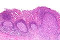
Diversion proctitis - low mag. (WC/Nephron)
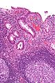
Diversion proctitis - high mag. (WC/Nephron)

Sign out
SIGMOID COLON, BIOPSIES:
- MILD ACTIVE COLITIS WITH LAMINA PROPRIA FIBROSIS, SEE COMMENT.
- NEGATIVE FOR DYSPLASIA.

COMMENT:
No granulomas are identified.  Follicular lymphoid hyperplasia is not identified;
however, there is no definite submucosa present.

Diverted segments of bowel can have inflammatory bowel disease-like changes.

In the context of a diverted segment of bowel, the findings are compatible with
a diversion colitis.
RECTUM, BIOPSY:
- CHRONIC ACTIVE PROCTITIS WITH FOCAL ULCERATION, CRYPTITIS AND CRYPT ABSCESSES.
- GRANULATION TISSUE.
- NEGATIVE FOR DYSPLASIA.

COMMENT:
No lymphoid hyperplasia is present. A small lymphoid aggregate is present.
Architectural distortion is present.

In the context of a diverted segment of bowel, the findings are compatible with
a diversion colitis.
See also
References
- ↑ 1.0 1.1 Edwards, CM.; George, B.; Warren, B. (Jan 1999). "Diversion colitis--new light through old windows.". Histopathology 34 (1): 1-5. PMID 9934577.
- ↑ Yeong, ML.; Bethwaite, PB.; Prasad, J.; Isbister, WH. (Jul 1991). "Lymphoid follicular hyperplasia--a distinctive feature of diversion colitis.". Histopathology 19 (1): 55-61. PMID 1916687.
- ↑ Ma, CK.; Gottlieb, C.; Haas, PA. (Apr 1990). "Diversion colitis: a clinicopathologic study of 21 cases.". Hum Pathol 21 (4): 429-36. PMID 2318485.
- ↑ Yantiss, RK.; Odze, RD. (Jan 2006). "Diagnostic difficulties in inflammatory bowel disease pathology.". Histopathology 48 (2): 116-32. doi:10.1111/j.1365-2559.2005.02248.x. PMID 16405661.
- ↑ Thorsen, AJ. (Feb 2007). "Noninfectious colitides: collagenous colitis, lymphocytic colitis, diversion colitis, and chemically induced colitis.". Clin Colon Rectal Surg 20 (1): 47-57. doi:10.1055/s-2007-970200. PMC 2780148. PMID 20011361. http://www.ncbi.nlm.nih.gov/pmc/articles/PMC2780148/.