Dermal cysts
Dermal cysts, also skin cysts, are common in dermatopathology. Dermatopathologists can diagnose 'em.
Overview
Common types:[1]
- Epidermal cyst (sebaceous cyst) -- most common.
- Pilar (trichilemmal) cyst.
- Dermoid cyst.
- Ganglion cyst.
- Milicem.
Epidermal necrosis
- This may be cystic. It is covered in the epidermal necrosis article, which covers erythema multiforme, Steven-Johnson syndrome and toxic epidermal necrolysis.
Common cysts
Venous lake
General
- Dilated vein.
Clinical:
- Blanch with pressure.[2]
Gross
- Purple/blue spot.
Images:
Microscopic
Features:[4]
- Lined by endothelium.
- Blood in lumen.
- +/-Fibrin in lumen.
- +/-Solar elastosis - very common.[5]
DDx:
- Angiokeratoma.
- Ectatic superficial dermal vessels.
- Irregular acanthosis.
- Longer rete ridges.
- Cherry hemangioma.[5]
Images:
- Venous lake (jhmi.edu).[3]
- Venous lake (dermpedia.org).[6]
- Venous lake (surgical pathologyatlas.com).
Sign out
SKIN LESION, RIGHT CHEEK, BIOPSY:
- VENOUS LAKE.
- SOLAR ELASTOSIS.
- NEGATIVE FOR NEVUS.
Epidermal inclusion cyst
Main article: Epidermal inclusion cyst
Pilar cyst
- AKA trichilemmal cyst.
Main article: Pilar cyst
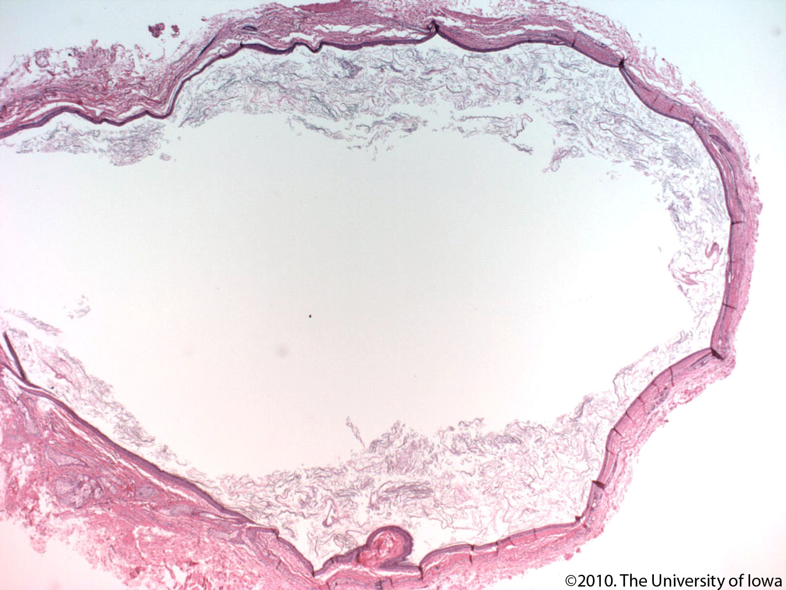
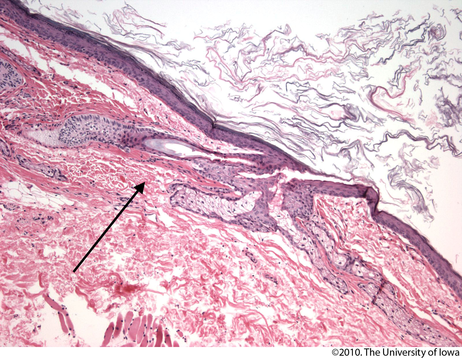
Dermoid cyst
General
- Benign.
- Congenital choristomas.[7]
- May be found in the ovary.
Microscopic
- Cyst lined by normal (keratinized) skin with adnexal structure (hair follicles, sweat glands, sebaceous glands).
DDx:
- Epidermal cyst - no adnexal structures.
Images:
Sign out
SKIN CYST, RIGHT LATERAL ORBIT, EXCISION:
- DERMOID CYST
- NEGATIVE FOR MALIGNANCY.
Pilonidal cyst
Main article: Pilonidal sinus
Less common
Steatocystoma
Main article: Steatocystoma
Digital mucous cyst
General
- Dome-shaped papule.
Microscopic
Features:[10]
- Mucous in superficial dermis - key feature.
- No epithelial lining; it is a pseudocyst.
Note:
- Mucin = glycolated proteins; may be part of mucous.
- Mucous = slippery secretion.
DDx:
Images:
Sign out
LESION, LEFT INDEX FINGER, EXCISION:
- DIGITAL MUCOUS CYST.
Apocrine cystadenoma
General
- Uncommon.
Microscopic
Features:[14]
- Multiloculated.
- Apocrine differentiation: columnar epithelium +/- apical snouts.
- Solid areas of epithelial proliferation.
- Papillary projections into the cyst.
Images:
See also
References
- ↑ Greenwald, J.; Heng, M. (2007). Toronto Notes for Medical Students 2007 (2007 ed.). The Toronto Notes Inc. for Medical Students Inc.. pp. D5. ISBN 978-0968592878.
- ↑ URL: http://dermatlas.med.jhmi.edu/derm/IndexDisplay.cfm?ImageID=-969536424. Accessed on: 13 August 2012.
- ↑ 3.0 3.1 3.2 URL: http://dermatlas.med.jhmi.edu/derm/result.cfm?Diagnosis=605386295. Accessed on: 13 August 2012.
- ↑ Weedon's Skin Pathology. 3rd Ed. P.895.
- ↑ 5.0 5.1 Busam, Klaus J. (2009). Dermatopathology: A Volume in the Foundations in Diagnostic Pathology Series (1st ed.). Saunders. pp. 551. ISBN 978-0443066542.
- ↑ URL: http://www.dermpedia.org/case/70-year-old-woman-with-nose-lesion. Accessed on: 21 June 2013.
- ↑ 7.0 7.1 7.2 Gandhi N, Syed NA, Alen R. Dermoid Cyst. EyeRounds.org. posted July 26, 2010; Available from: http://www.EyeRounds.org/cases/115-dermoid-cyst.htm. Accessed on: 22 September 2011.
- ↑ Mitchell, Richard; Kumar, Vinay; Fausto, Nelson; Abbas, Abul K.; Aster, Jon (2011). Pocket Companion to Robbins & Cotran Pathologic Basis of Disease (8th ed.). Elsevier Saunders. pp. 596. ISBN 978-1416054542.
- ↑ URL: http://emedicine.medscape.com/article/788127-overview. Accessed on: 10 September 2012.
- ↑ 10.0 10.1 10.2 10.3 URL: http://www.dermpedia.org/dermpedia-textbook/digital-mucous-myxoid-cyst. Accessed on: 17 January 2012.
- ↑ URL: http://dictionary.reference.com/browse/mucous. Accessed on: 8 January 2012.
- ↑ URL: http://dictionary.reference.com/browse/mucus. Accessed on: 8 January 2012.
- ↑ URL: http://www.dermpedia.org/case/digital-mucous-cyst-ganglion-type. Accessed on: 5 July 2013.
- ↑ Busam, Klaus J. (2009). Dermatopathology: A Volume in the Foundations in Diagnostic Pathology Series (1st ed.). Saunders. pp. 316. ISBN 978-0443066542.