Course:Introduction to Neuropathology
Course Neuropathology is a online collection of images and descriptions of specimens used for teaching medical students and residents.
This page is divided into following courses:
- Basic neuropathology - preclinical medical education
- Molecular neuropathology - ideal for bachelor of molecular medicine or oncology
- Advanced neuropathology - clinical medical education
Basic neuropathology
Day one
Meningioma
Picture 1

Picture 2
Picture 3
Picture 4
Picture 5


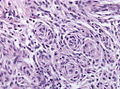
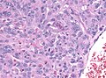
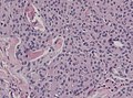
This H&E stain displays parts of a moderately cellular tumor growing with ovoid elongated nuclei (Pictures 1+2). There are no clear-cut cell borders discernible in light microscopy. This interveawing is called a syncytium. WHO Grading of the tumour is dependent of the mitotic activity (Picture 3) or histological hallmarks such as prominent nucleoli, high nuclear to cytoplasmic ratio, CNS infiltation etc.. Focal nuclear clearing (Nuclear pseudoinclusions, Picture 4) are typical cut phenomenon. The round calcified inclusions (Psammoma bodies) are characteristic for a meningioma.
Other language: German
|
|---|
|
Das H&E-Präparat zeigt Anteile eines mäßig zelldichten in Zügen und Wirbeln orientierten Tumors mit ovoid elongierten Tumorzellkernen (Bilder 1+2). Eindeutige Zellgrenzen lassen sich lichtmikroskopisch nicht definieren, was als pseudosynzytialer Aspekt bezeichnet wird. Die Wertung des Tumors nach WHO ist abhängig von der mitotischen Aktivität (Bild 3), oder durch histologische Kriterien wie z.B. prominente Nukleolen, Kern-Plasma-Relation, ZNS-Infiltration etc.. Lochkernzellen (Bild 4) sind hingegen ein typisches Anschnittphänomen. Auch rundliche Verkalkungen (Bild 5), sogenannte Psammomkörper sind charakteristisch für Meningeome. |
Astrocytoma
Picture 1
Picture 2
Picture 3


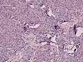
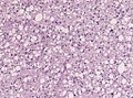
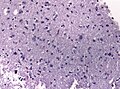
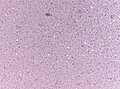
This specimen contains fragments of a diffusely growing tumor with only slight inclreased cell density and focally microcysts withing the neuropil background (Picture 1). Although many of the astrocytic tumour cells look quite similiar there is increased pleomorphism, mostly larger and a more dense chromatin. There are no mitoses seen in this tumour (Picture 2). The tumour borders are not clear, there is just a decrease of cell density a the tumor infiltration zone. In this area one is not always sure which cells are still neoplastic and which cells are normal or reactive astrocytes of the normal brain (Picture 3).
Other language: German
|
|---|
|
Das Präparat besteht aus Fragmenten eines diffus wachsenden, gering zelldichten Tumors, welcher fokal mikrozystische Auflockerungen aufweist (Bild 1). Trotz hoher Ähnlichkeit der astrozytären Tumorzellen finden sich fokal vermehrte Pleomorphie der Kerne, diese meist etwas größer und chromatindichter. Mitosen finden sich in diesem Tumor nicht. (Bild 2). Die Tumorgrenzen sind unscharf, man beobachtet lediglich eine diffus abnehmende Zelldichte im Bereich der Infiltrationszone, in der man nicht genau sagen kann, welches noch Tumorzellen sind und welche bereits normale bzw. reaktiv veränderte Astrozyten darstellen (Bild 3). |