Difference between revisions of "Columnar cell change of the breast"
Jump to navigation
Jump to search
(Created page with "'''Columnar cell change of the breast''', usually '''columnar cell change''' (abbreviated '''CCC'''), is a benign finding in breast pathology. It is also known as '''blun...") |
|||
| Line 4: | Line 4: | ||
==General== | ==General== | ||
*Columnar cell change is associated with (benign) [[breast calcification|calcification]] - '''key point'''. | *Columnar cell change is associated with (benign) [[breast calcification|calcification]]<ref name=pmid19089736>{{Cite journal | last1 = Jara-Lazaro | first1 = AR. | last2 = Tse | first2 = GM. | last3 = Tan | first3 = PH. | title = Columnar cell lesions of the breast: an update and significance on core biopsy. | journal = Pathology | volume = 41 | issue = 1 | pages = 18-27 | month = Jan | year = 2009 | doi = 10.1080/00313020802563486 | PMID = 19089736 }}</ref> - '''key point'''. | ||
==Microscopic== | ==Microscopic== | ||
Revision as of 04:50, 1 July 2016
Columnar cell change of the breast, usually columnar cell change (abbreviated CCC), is a benign finding in breast pathology.
It is also known as blunt duct adenosis.
General
- Columnar cell change is associated with (benign) calcification[1] - key point.
Microscopic
Features:
- Secretory cells (line gland lumen) have columnar morphology.
- May have "apical snouts".
- Blebs or round balls eosinophilic material appear to be adjacent to the cell at their luminal surface.
- The snouts are attached to the cell-- appear as round ball only in the plane of section.
- Cytoplasm +/-eosinophilia.
- Often purple luminal calcifications
DDx:
- Flat epithelial atypia (>2 cell layers).[citation needed]
- If the columnar cells shows low to intermediate grade atypia the process is termed "flat epithelial atypia"
- If higher grade atyia is present the lesion is termed "flat DCIS" (clinging carcinoma)
Images

CCC - intermed. mag.
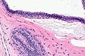
CCC - high mag.
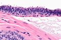
CCC - very high mag.


www=
Sign out
- Usually not reported.
See also
References
- ↑ Jara-Lazaro, AR.; Tse, GM.; Tan, PH. (Jan 2009). "Columnar cell lesions of the breast: an update and significance on core biopsy.". Pathology 41 (1): 18-27. doi:10.1080/00313020802563486. PMID 19089736.