Difference between revisions of "Choroid plexus papilloma"
Jump to navigation
Jump to search
(tweak) |
(tweak some more) |
||
| Line 7: | Line 7: | ||
| Micro = papillary epithelial structures with bland cytology, mitoses uncommon | | Micro = papillary epithelial structures with bland cytology, mitoses uncommon | ||
| Subtypes = | | Subtypes = | ||
| LMDDx = metastatic papillary [[adenocarcinoma]], [[ELST]] | | LMDDx = [[choroid plexus carcinoma]], metastatic papillary [[adenocarcinoma]], [[ELST]] | ||
| Stains = | | Stains = | ||
| IHC = Kir 7.1 +ve, EAAT1 +ve, GFAP +ve/-ve, S-100 +ve/-ve, CK +ve/-ve | | IHC = Kir 7.1 +ve, EAAT1 +ve, GFAP +ve/-ve, S-100 +ve/-ve, CK +ve/-ve | ||
| Line 61: | Line 61: | ||
* Atypical CPP: 2 or more mitoses /10 HPF.<ref name=pmid19543851>{{Cite journal | last1 = Wrede | first1 = B. | last2 = Hasselblatt | first2 = M. | last3 = Peters | first3 = O. | last4 = Thall | first4 = PF. | last5 = Kutluk | first5 = T. | last6 = Moghrabi | first6 = A. | last7 = Mahajan | first7 = A. | last8 = Rutkowski | first8 = S. | last9 = Diez | first9 = B. | title = Atypical choroid plexus papilloma: clinical experience in the CPT-SIOP-2000 study. | journal = J Neurooncol | volume = 95 | issue = 3 | pages = 383-92 | month = Dec | year = 2009 | doi = 10.1007/s11060-009-9936-y | PMID = 19543851 }}</ref> | * Atypical CPP: 2 or more mitoses /10 HPF.<ref name=pmid19543851>{{Cite journal | last1 = Wrede | first1 = B. | last2 = Hasselblatt | first2 = M. | last3 = Peters | first3 = O. | last4 = Thall | first4 = PF. | last5 = Kutluk | first5 = T. | last6 = Moghrabi | first6 = A. | last7 = Mahajan | first7 = A. | last8 = Rutkowski | first8 = S. | last9 = Diez | first9 = B. | title = Atypical choroid plexus papilloma: clinical experience in the CPT-SIOP-2000 study. | journal = J Neurooncol | volume = 95 | issue = 3 | pages = 383-92 | month = Dec | year = 2009 | doi = 10.1007/s11060-009-9936-y | PMID = 19543851 }}</ref> | ||
DDx: | |||
* | *[[Choroid plexus carcinoma]]. | ||
* | *[[Metastatic]] papillary carcinoma. | ||
* | *[[Endolymphatic sac tumour]] (ELST). | ||
===Images=== | ===Images=== | ||
| Line 79: | Line 75: | ||
www: | www: | ||
*[http://path.upmc.edu/cases/case551.html Choroid plexus papilloma - oncocytic variant - several images (upmc.edu)]. | *[http://path.upmc.edu/cases/case551.html Choroid plexus papilloma - oncocytic variant - several images (upmc.edu)]. | ||
==IHC== | |||
*CK7 +ve. | |||
*CK20 -ve (typically). | |||
*S-100 +ve/-ve. | |||
*EMA -ve. | |||
*GFAP +ve/-ve. | |||
*Kir 7.1 +ve. | |||
*EAAT1 +ve. | |||
==See also== | ==See also== | ||
Revision as of 07:11, 1 May 2015
| Choroid plexus papilloma | |
|---|---|
| Diagnosis in short | |
 Choroid plexus papilloma H&E stain. | |
|
| |
| LM | papillary epithelial structures with bland cytology, mitoses uncommon |
| LM DDx | choroid plexus carcinoma, metastatic papillary adenocarcinoma, ELST |
| IHC | Kir 7.1 +ve, EAAT1 +ve, GFAP +ve/-ve, S-100 +ve/-ve, CK +ve/-ve |
| Site | all ventricles, cerebellopontine angle - see brain tumours |
|
| |
| Clinical history | children |
| Symptoms | hydrocephalus |
Choroid plexus papilloma, abbreviated CPP, is a benign neuropathology arising in the ventricles of the brain.
General
Features: [1]
- Low grade
- WHO grade I (ICD-O: 9390/1).
- WHO grade II - atypical choroid plexus papilloma (ICD-O: 9390/1).
- Less than 1% of all brain tumours.
- Approximately 10-20% of all tumours in the first year of life.
- Congenital tumours reported.
- Usually laternal ventricle in children.[2]
Gross
Features: [1]
- Adhere to ventricular walls.
- Circumscribed.
- Villous architecture.
- 50% lateral ventricles.
- 5% third ventricle.
- 40% fourth ventricle.
- 5% multiple tumours or cerebellopontine angle.
Microscopic
Features: [3]
- Simple epithelium.
- Papillae.
- Psammoma bodies.
- Rarely oncocytic changes, mucinous degeneration.
- Necrosis and brain invasion is uncommon.
- Atypical CPP: 2 or more mitoses /10 HPF.[4]
DDx:
- Choroid plexus carcinoma.
- Metastatic papillary carcinoma.
- Endolymphatic sac tumour (ELST).
Images
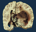
Choroid plexus papilloma can become quite large. (WC/marvin101)
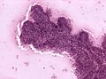
Intraoperative smear of a choroid plexus papilloma. (WC/jensflorian)
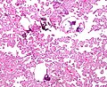
Choroid plexus papilloma. (WC)
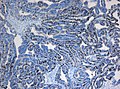
Elevated proliferative activity (MIB-1) in atypical CPP. (WC/marvin101)


www:
IHC
- CK7 +ve.
- CK20 -ve (typically).
- S-100 +ve/-ve.
- EMA -ve.
- GFAP +ve/-ve.
- Kir 7.1 +ve.
- EAAT1 +ve.
See also
References
- ↑ 1.0 1.1 The International Agency for Research on Cancer (Editors: Louis, D.N.; Ohgaki, H.; Wiestler, O.D.; Cavenee, W.K.) (2007). Pathology and Genetics of Tumours of Tumors of the Central Nervous System (IARC WHO Classification of Tumours) (4th ed.). Lyon: World Health Organization. pp. 82. doi:10.1007/s00401-007-0243-4. ISBN 978-9283224303.
- ↑ URL: http://emedicine.medscape.com/article/250795-overview. Accessed on: 3 June 2011.
- ↑ Menon, G.; Nair, SN.; Baldawa, SS.; Rao, RB.; Krishnakumar, KP.; Gopalakrishnan, CV.. "Choroid plexus tumors: an institutional series of 25 patients.". Neurol India 58 (3): 429-35. doi:10.4103/0028-3886.66455. PMID 20644273.
- ↑ Wrede, B.; Hasselblatt, M.; Peters, O.; Thall, PF.; Kutluk, T.; Moghrabi, A.; Mahajan, A.; Rutkowski, S. et al. (Dec 2009). "Atypical choroid plexus papilloma: clinical experience in the CPT-SIOP-2000 study.". J Neurooncol 95 (3): 383-92. doi:10.1007/s11060-009-9936-y. PMID 19543851.