Choroid plexus carcinoma
Jump to navigation
Jump to search
| Choroid plexus carcinoma | |
|---|---|
| Diagnosis in short | |
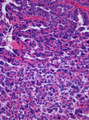
 Choroid plexus carcinoma. H&E stain. | |
|
| |
| LM | choroid plexus epithelium with nuclear pleomorphism & high NC ratio, mitoses, necrosis, +/-brain invasion |
| LM DDx | choroid plexus papilloma, atypical plexus papilloma, atypical teratoid/rhabdoid tumour |
| IHC | cytokeratins +ve, EMA -ve, INI1 +ve, Ki-67 high |
| Site | brain - intraventricular mass, classically posterior fossa |
|
| |
| Clinical history | typically children |
| Prevalence | uncommon |
Choroid plexus carcinoma is an uncommon neuropathology tumour and the malignant counter part of choroid plexus papilloma.
General
- Usually pediatric population.
- Malignant counterpart of choroid plexus papilloma.[1]
- Poor prognosis - WHO grade III.[2]
- Classically posterior fossa.
- Intraventricular mass.
Microscopic
Features:[1]
- Choroid plexus epithelium with nuclear pleomorphism & high NC ratio.
- Mitoses.
- Necrosis.
- +/-Brain invasion.
DDx:
- Choroid plexus papilloma.
- Atypical plexus papilloma - has features intermediate between choroid plexus papilloma and choroid plexus carcinoma.[2]
- Atypical teratoid/rhabdoid tumour.
Images
CPC. (WC/Martin Hasselblatt)
www:
IHC
Features:[2]
- Cytokeratins +ve.
- EMA usu. -ve.
- GFAP -ve (~20% +ve).
- Ki-67 high.
- Useful to diff. from benign counterpart.
- INI1 +ve.
See also
References
- ↑ 1.0 1.1 Singh, A.; Vermani, S.; Shruti, S.. "Choroid plexus carcinoma: report of two cases.". Indian J Pathol Microbiol 52 (3): 405-7. doi:10.4103/0377-4929.55009. PMID 19679976.
- ↑ 2.0 2.1 2.2 Menon, G.; Nair, SN.; Baldawa, SS.; Rao, RB.; Krishnakumar, KP.; Gopalakrishnan, CV.. "Choroid plexus tumors: an institutional series of 25 patients.". Neurol India 58 (3): 429-35. doi:10.4103/0028-3886.66455. PMID 20644273.