Difference between revisions of "Avascular necrosis of the femoral head"
Jump to navigation
Jump to search
(split out) |
|||
| Line 23: | Line 23: | ||
</ref> | </ref> | ||
*Empty lacunae (indicative of necrotic bone). | *Empty lacunae (indicative of necrotic bone). | ||
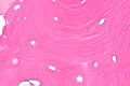
===Images=== | |||
<gallery> | |||
Image: Necrotic bone -- intermed mag.jpg | AVN - intermed. mag. (WC/Nephron) | |||
Image: Necrotic bone -- high mag.jpg | AVN - high mag. (WC/Nephron) | |||
Image: Necrotic bone -- very high mag.jpg | AVN - very high mag. (WC/Nephron) | |||
</gallery> | |||
==Sign out== | ==Sign out== | ||
Revision as of 02:16, 20 December 2013
Avascular necrosis of the femoral head is necrosis of the head of the femur to the vascular compromise.
It is often just referred to as avascular necrosis, abbreviated AVN.
General
Risk factors:
- Oral steroids, e.g. prednisone.[1]
- Cushing disease.
- Cushing syndrome.
- Radiation.
Gross
Features:[2]
- Wedge-shaped pale yellow abnormality below cartilage.
- +/-Cartilage separates from the bone.
- +/-Deformation of femoral head.
Image:
Microscopic
Features:[3]
- Empty lacunae (indicative of necrotic bone).
Images
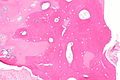
AVN - intermed. mag. (WC/Nephron)

AVN - high mag. (WC/Nephron)
AVN - very high mag. (WC/Nephron)


Sign out
FEMORAL HEAD, RIGHT, HIP ARTHROPLASTY:
- AVASCULAR NECROSIS OF THE FEMORAL HEAD.
AVN and degenerative joint disease
FEMORAL HEAD AND JOINT CAPSULE, LEFT, HIP ARTHROPLASTY:
- AVASCULAR NECROSIS OF THE FEMORAL HEAD.
- DEGENERATIVE JOINT DISEASE WITH MILD SYNOVITIS AND VILLOUS HYPERPLASIA.
- NEGATIVE FOR MALIGNANCY.
FEMORAL HEAD AND JOINT CAPSULE, RIGHT, HIP ARTHROPLASTY:
- AVASCULAR NECROSIS OF THE FEMORAL HEAD.
- DEGENERATIVE JOINT DISEASE.
- BENIGN JOINT CAPSULE TISSUE.
- NEGATIVE FOR MALIGNANCY.
Remote AVN
FEMORAL HEAD AND JOINT CAPSULE, LEFT, HIP ARTHROPLASTY:
- FEMORAL HEAD WITH DEGENERATIVE JOINT DISEASE AND MARKED DEFORMATION CONSISTENT WITH A HISTORY OF
AVASCULAR NECROSIS.
- JOINT CAPSULE WITH MINIMAL CHRONIC INFLAMMATION.
Micro
The sections show a femoral head with loss of cartilage and focal vertical cleft formation in the remaining thinned cartilage. Subchondral sclerosis is present. The underlying bone is viable. Bone marrow is present. The red blood cells have a sickled morphology.
Joint capsule tissue with focal lymphocytes and plasma cells is present.
See also
References
- ↑ URL: http://www.merckmanuals.com/professional/musculoskeletal_and_connective_tissue_disorders/osteonecrosis/osteonecrosis.html. Accessed on: 30 April 2012.
- ↑ Lester, Susan Carole (2005). Manual of Surgical Pathology (2nd ed.). Saunders. pp. 224. ISBN 978-0443066450.
- ↑ Steffen, RT.; Athanasou, NA.; Gill, HS.; Murray, DW. (Jun 2010). "Avascular necrosis associated with fracture of the femoral neck after hip resurfacing: histological assessment of femoral bone from retrieval specimens.". J Bone Joint Surg Br 92 (6): 787-93. doi:10.1302/0301-620X.92B6.23377. PMID 20513874.