Difference between revisions of "Autopsy"
m (→Respiratory system: sm fix) |
|||
| Line 306: | Line 306: | ||
==Skull & brain== | ==Skull & brain== | ||
===Opening the skull=== | |||
#One should saw through the skull completely, i.e. one should not "crack" the skull open with a chisel. | #One should saw through the skull completely, i.e. one should not "crack" the skull open with a chisel. | ||
#*Cracking open the skull may result in artefactual fractures that are impossible to differentiate from antemortem fractures. | #*Cracking open the skull may result in artefactual fractures that are impossible to differentiate from antemortem fractures. | ||
#*Cuts into the brain (from opening the skull) are not difficult to distinguish from antemortem injuries. | #*Cuts into the brain (from opening the skull) are not difficult to distinguish from antemortem injuries. | ||
===Extraction of the brain=== | |||
#Cranial nerves should be cut. | #Cranial nerves should be cut. | ||
#Tentorium should be released. | #Tentorium should be released. | ||
| Line 320: | Line 322: | ||
===Brain=== | ===Brain=== | ||
==== | Retension of the brain (for prolonged fixation): | ||
*Survival interval in hospital. | |||
*Neurosurgical intervention. | |||
*Clinical Dx of DAI. | |||
*Hypoxic changes (make brain hard to cut). | |||
====Sectioning==== | |||
Up-down anatomically oriented (right side-up): | Up-down anatomically oriented (right side-up): | ||
#Examine meninges. | #Examine meninges. | ||
Revision as of 04:58, 18 October 2010
Autopsy is a part of pathology.
In a hospital autopsy the most important thing is: proper consent.
Consent
- Consent should be given by the executer of the estate.[1]
If the executer of the estate is not specified the hierarchy is as follows:
- Spouse - by marriage (same sex or opposite sex) or by common-law or by together the parents of a child or by cohabitation agreement (in law).
- If no spouse, any children 16+ years old,
- If no children, either parent,
- If no parent, any brother or sister 16+ years old,
- If no sibling, any next-of-kin 16+ years old,
- If no next-of-kin, the person lawfully in possession of the body (not the hospital).
Notes:
- The power a person that is designated as power of attorney for health care decisions does not have the authority to consent for an autopsy; their power ends with death (unless they are also the executer of the estate).
- In clinical medicine, it is allowable to skip down the hierarchy if the "consent giver" is not reachable, e.g. if a child of the patient is present they can consent in emergency circumstances, if the spouse is not reachable. In the context of autopsies, the hierarchy has to be followed strictly, as there is no such thing as an "emergency autopsy"; it is not acceptable to ask the child of the decedent 'cause they aren't distraught like the spouse of the decedent.
Religious objections
- There are religious objections to autopsy among Jews and Muslims.[2][3]
- It is not considered good practise to agree to restrictions that will impair a complete assessment, e.g. "stop as soon as one has the cause of death", especially in the medicolegal context (when the extent of the autopsy is at the pathologist's discretion). It is often said that... incomplete autopsies give incomplete answers.
External exam
General
- Very important in the forensic context.
- Medical devices, tubes and lines should be left in situ to allow the determination of their precise location within the body;[4] it is very difficult to determine what the location of a line was once it is removed.
Extremities
- Fingers should be identified by name (e.g. ring finger), as some people number the digits 1-4 and consider the thumb separately, while others number 'em 1-5.[5]
Body should be examined for defensive-type wounds:
- Between the finger, esp. thumb and pointing finger.
- Dorsal aspect of the hand.
- Forearm.
Findings
- External exam findings are found in the forensic pathology article.
Internal exam
General
- This is usually where the money is in hospital autopsies.
- Like surgeons say... you should never cut anything until you're sure what it is.
Before the first incision
If there is suspicion of pneumothorax - one the the three following can be done:[6]
- Create a "pleural window" (between ribs by removing soft tissue... without entering the pleural cavity).
- Open chest underwater and watch for air bubbles.
- Needle puncture with water filled syringe - where plunger has been removed.
- If there are neck abnormalities or suspicion of pressure on the neck, it is prudent to remove the cranial contents and thoracic contents before doing the neck dissection.[7]
- If the above does not apply, i.e. there is no neck injury/suspected neck injury, the tongue & neck can be taken together with the thorax pluck or organ pluck.
Incisions
- Y-shaped incision (standard):
- Superior "points" of the Y ~ at the deltoid muscle.
- "Confuence of lines" in the Y ~ at the suprasternal notch.
- Neck dissection incisions:
- Deltoid to mastoid process.
Sternum
- Usually of no interest.
- May have cleft or foramen - as an anatomical variant.[8]
Blood clots - pre- and post-mortem
| Feature/time | Pre-mortem | Post-mortem |
| Shininess | dull | shiny |
| Adherent to wall | yes[9] | no |
| Colour | grey; may have zebraic appearance (lines of Zahn) - red alt. with grey/yellow |
dark purple or bilayered yellow/red |
| Pressurized | yes; "ejects itself" from lumen | no; needs to be pulled-out |
| Consistency -elastic modulus (E) -fracture toughness (K) |
firm (high E) brittle (low K) |
jello (low E) elastic (high K) |
| Image - gross | thrombus (pathguy.com), thrombus (thrombosisadviser.com) |
coronary thrombus (luc.edu)[10] |
| Image - micro. | pre- & post-mortem (elsevier.es)[11] | thrombus (oxfordjournals.org), thrombi (ucsf.edu) |
Notes:
- Post-mortem thrombi: one (superior) yellow portion (called "chicken fat") and one (dependent) red portion (RBCs); components layer due to gravity.
Retrieval of the (organ) "pluck"
One piece (standard):
- Diaphragm cut: at chest wall.
- Superior cut: through arch vessels.
- Inferior cut: through the common iliac vessels.
- Posterior:
- Thorax: cut paravertebral.
- Abdomen: blunt dissection for kidneys, then paravertebral.
Two pieces (thorax & abdomen):
- May be preferred in a "glued" (fibrosed/post-surgical) abdomen.
- Procedure - similar to one piece but:
- Cut aorta distal to the left subclavian + detach from thoracic pluck.
- Cut esophagus distal to pharynx + detach from thoracic pluck.
Neck organ pluck
- Trim tissue posterior to horns of the thyroid.
- Cut between thyroid horns & hyoid.
- Cut off base of tongue.
Hyoid bone
- Important in forensic pathology.
- Fracture is seen in manual strangulation.
- May appear fractured if triticeous cartilage (or triticeal cartilage) is present;[12][13] triticeous cartilage may be confused with a fragment of hyoid bone.
- Triticeous is pronounced tri-tish´us.[14]
Dissection of thoraco-abdominal pluck
Process
- Place organ pluck with anterior aspect down.
- Open aorta (posterior aspect - from just distal to subclavian + common iliacs).
- Probe celiac trunk, SMA, renal arteries, IMA.
- Open renal arteries.
- Blunt dissection to separate aorta from thorax distal to left subclavian artery.
- Cut through aorta distal to left subclavian artery.
- Separate aorta from pluck.
- Open IVC to diaphragm.
- Check renal veins.
- Separate IVC.
- Take down esophagus using blunt dissection (to separate from thorax).
- Separate thorax and abdomen - by dissecting through pleural adhesions, cut through pericardium.
Common finding(s)
- Aorta = atheroscerlosis.
Things to think about
- Renal arteries = atherosclerosis (old), fibromuscular dysplasia (young).
- Renal veins = renal cell carcinoma.
Thoracic pluck
Heart
Respiratory system
- Open trachea + bronchus (see Note).
- Examine proximal airway.
- Examine proximal pulmonary arteries.
- Slice lungs - one lobe at a time (easier to cut).
- Squeeze lungs - to test for the presence of pus and edema fluid.[16]
Note:
- If the lungs are to be inflated the bronchi should be left long.
Common findings
- Plural adhesions.
- Plural effusions.
- Edema - fluid comes-out when you squeeze 'em, "heavy" (large mass).
- Emphysematous change (usu. upper lung zone predominant) +/- black pigment (anthracosis).
- Consolidation - best appreciated by running a finger over the cut surface of the lung with a small-to-moderate amount of pressure.
Abdominal organ pluck
Adrenal glands
- Place cuts at anatomical location.
- Take section for stock.
Common findings
- Cortical adenomas - seen in ~ 2% of autopsies.[17]
- Metastatic cancer, esp. in the context of lung cancer.
Others
- Atrophy.
- Hyperplasia (bilateral).
- Hemorrhage (Waterhouse-Friderichsen syndrome).
Stomach
- Opened along greater curvature.
- GE junction should not be opened if portal hypertension is suspected (see: esophagus).
Findings
- Haemorrhage = usu. post-mortem.
- Small ~1 mm pellets (medicinal capsule contents).
Esophagus
- Should be everted, if portal hypertension is suspected, as varices are thus more readily demonstrated.[18]
- Stomach opened (without opening GE junction).
- String tied to proximal esophagus.
- Forceps inserted from stomach to grasp tied end and invert esophagus.
Omentum
- It is a good idea to trim this from the stomach.
Spleen
- Separate stomach from spleen.
- Identify the splenic vessels.
- To separate the spleen from the pluck:
- Cut across vessels at splenic hilum - close to the spleen.
Common findings
- Sugar-coated spleen.
- Properly referred to as hyaloserositis of the spleen.
- Capsule of the spleen is white - resembles sugar-coating.
- Importance: none - benign.
- Splenomegaly secondary to portal hypertension.
Kidney
- Renal vein & artery already examined.
- +/-Open ureter.
- Strip capsule.
- Left ureter is left long - to tell apart the kidneys.
- Kidney (nearly) bisected in coronal plane from anti-ureteric surface; a small rim of tissue is left on the hilar (or ureteric) aspect -- to keep the kidney as one piece.[19]
Common pathologic findings
- Size of the kidney - small kidneys are seen in chronic renal failure.
- Nephrosclerosis:
- Flea-bitten appearance - seen in hypertension.[20]
- Acute tubular necrosis (ATN).
- ATN is difficult (or impossible) to prove on autopsy material.
- Look for:
- Heme-granular casts in the lumen.
- Regenerative activity (mitoses).
Liver
- Remove diaphragm (with scissors).
- Separated liver from abdominal pluck at porta hepatis.
- Open Gallbladder.
- Get mass.
- Slice axially - as seen on CT imaging.
Notes:
- If there is a suspected biliary tract obstruction:
- Open the duodenum (from distal end) and identify the duodenal papilla - this is usually obvious as everything distal to the duodenal papilla is usually light brown (bile-stained).
- Compress the gallbladder and bile should emerge from the duodenal papilla.
- Disect bilary tree from duodenal papilla to the porta hepatis.
Common pathologic findings
Liver
- Firm - suggests cirrhosis.
- Micronodularity (with uniformity of the nodules) - suggestive of alcoholic cirrhosis.
- Macronodularity (with heterogeneity of the nodules) - suggestive of viral hepatitis.
- Yellow or pale liver:
- DDx:
- Shock liver.
- Steatosis or steatohepatitis.
- DDx:
- Congested (blood red/yellow) "nutmeg liver".
Gallbladder
- Gall stones.
- Mucosa with strawberry-like appearance (cholesterolosis).
Pancreas
- Serially section along axis of pancreatic duct.
- Pieces of head & tail for stock or stock and histology.
Genitourinary-rectal or pelvic pluck
Female[21]
- Orient rectum down, bladder up.
- Open bladder with scissors via urethra with cuts in sagittal plane.
- Flip over specimen.
- Open rectum with scissors with cuts in the sagittal plane.
- Flip over specimen.
- Open vagina with cuts on its lateral aspect.
- Open cervix & uterus with cuts on its lateral aspect.
- Bisect ovaries.
Common pathologic findings
- Ovary:
- Corpus luteum - bright yellow (non-pathologic).
- Simple cyst.
- Urinary bladder:
- Focal haemorrhage secondary to urinary catheter.
Male
- Orient rectum down, bladder up.
- Open bladder with scissors via urethra/prostate with cuts in sagittal plane.
- Serially section prostate in the planes defined by the axis of the urethra.
- Flip over specimen.
- Open rectum with scissors with cuts in the sagittal plane.
Common pathologic findings
- Prostate gland:
- Enlargement (benign prostatic hyperplasia).
- Urinary bladder:
- Trabeculation secondary to BPH.
- Focal haemorrhage secondary to urinary catheter.
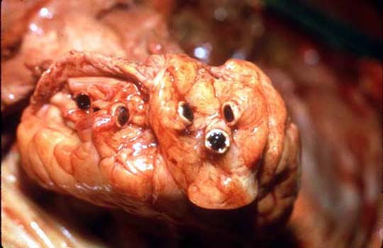
Skull & brain
Opening the skull
- One should saw through the skull completely, i.e. one should not "crack" the skull open with a chisel.
- Cracking open the skull may result in artefactual fractures that are impossible to differentiate from antemortem fractures.
- Cuts into the brain (from opening the skull) are not difficult to distinguish from antemortem injuries.
Extraction of the brain
- Cranial nerves should be cut.
- Tentorium should be released.
- Spinal cord should be completely cut as far down as possible.
- Dura should be stripped from the skull base.
Notes:
- Scalp hematoma:
- May result from intracranial pressure.
Brain
Retension of the brain (for prolonged fixation):
- Survival interval in hospital.
- Neurosurgical intervention.
- Clinical Dx of DAI.
- Hypoxic changes (make brain hard to cut).
Sectioning
Up-down anatomically oriented (right side-up):
- Examine meninges.
- Remove dura.
- Examine venous sinuses.
- Look for gyri effacement - due to edema or haematomas.
Not up-down anatomically oriented (top down):
- Remove brain stem - with one cut ~ at level of mid-brain.
- Cerebrum section - first cut at mammillary bodies.
- Subsequent cuts ~ 1.0 cm in thickness.
Anatomic variants
- Metopic suture - midline in frontal bone.[22]
- Wormian bones = "extra" bone at the sutures in the skull.
Anatomy
- Lambdoidal suture - occipital bone/parietal bones.
- Coronal suture - frontal bone/parietal bone.
Weird stuff
- Leukostasis in acute myelogenous leukemia can lead to congestion of organs and fatal haemorrhages.
- Hyperviscosity syndrome - in leukemia.[25]
Starvation
- Serous fat atrophy.
- Gross appearance: brown goo replaces fat.
- May be associated with blood vessel tortuosity.[26]
- Gross appearance: brown goo replaces fat.
Normal organ masses
Caucasoid population of 684 adults:[27]
| Men | Women | |
| Heart | 365 +/- 71 g | 312 +/- 78 g |
| Right lung | 663 +/- 239 g | 546 +/- 207 g |
| Left lung | 583 +/- 216 g | 467 +/- 174 g |
| Liver | 1677 +/- 396 g | 1475 +/- 362 g |
| Spleen | 156 +/- 87 g | 140 +/- 78 g |
| Right kidney | 162 +/- 39 g | 135 +/- 39 g |
| Left kidney | 160 +/- 41 g | 136 +/- 37 g |
Histology
General
It's the at the pathologists discretion. In decomp cases it is reasonable to submit nothing.
Forensic context
A standard (minimum) for adult homicides is:
- Heart (1).
- Lung (1).
- Liver (1).
- Kidney (1).
Hospital autopsies
Non-neuro
- Thyroid gland (1).
- Lung - each lobe (5).
- Heart - posterior papillary muscle & LV, anterior papillary muscle & LV (2).
- More extensive histologic sectioning is discussed in the heart article.
- Liver - left lobe, right lobe (2).
- Spleen - including hilum & capsule (1).
- Adrenal gland (1).
- Gastrointestinal tract:
- The quality if often very poor (even in "fresh" bodies). These are often omitted.
- Pancreas - head & tail (1).
- Stomach (1).
- Esophagus (1).
- Small bowel (1).
- Large bowel (1).
- The quality if often very poor (even in "fresh" bodies). These are often omitted.
- Genitourinary tract:
- Kidney - each side (2).
- Gender specific:
- Male:
- Prostate gland (1).
- Testis (1).
- Female:
- Uterus +/- cervix (1).
- Ovary + tube (1).
- Male:
- Bone marrow (1).
Neuro
- Pituitary gland (1).
- Cortex usu. frontal/parietal (1).
- Hippocampus (1).
- Cerebellum (1).
- Brain stem (1).
- Basal ganglia (1).
Stock
- Thyroid gland.
- Heart:
- Complete (axial) slice (including RV and LV).
- Tissue with SA node.
- Tissue with AV node.
- Lung - all five lobes.
- Spleen.
- Urinary:
- Kidney.
- Urinary bladder.
- Gastrointestinal tract:
- GE junction.
- Stomach.
- Small bowel.
- Large bowel.
- Appendix.
- Pancreas (head & tail).
- Liver.
- Reproductive:
- Male:
- Prostate gland.
- Testis.
- Female:
- Breast.
- Uterus.
- Ovary.
- Male:
- Neurologic:
- Hippocampus + basal ganglia.
- Cortex.
- Brainstem.
- Cerebellum.
- Skin.
- Nerve.
- Muscle.
- Lymph nodes.
Negative autopsy
Definition:
- A negative autopsy is a post-mortem exam that has no anatomical or toxicological cause of death.
- This does not preclude the presence of pathology (that is not sufficient to cause death).
Cause of death (in a negative autopsy):
- Unascertained.
Considerations/Causes
- Channelopathy (unrecognized).
- Electrocution (unrecognized).
- Decomposition.
- Sudden Unexpected Death in Epilepsy (SUDEP)[28] - missing history.
See also
References
- ↑ URL: http://www.docstoc.com/docs/51609856/CONSENT-FOR-AUTOPSY. Accessed on: 27 September 2010.
- ↑ Burton, Julian L.; Rutty, Guy N. (2010). The Hospital Autopsy A Manual of Fundamental Autopsy Practice (3rd ed.). Oxford University Press. pp. 43. ISBN 978-0340965146.
- ↑ Burton, Julian L.; Rutty, Guy N. (2010). The Hospital Autopsy A Manual of Fundamental Autopsy Practice (3rd ed.). Oxford University Press. pp. 47. ISBN 978-0340965146.
- ↑ Burton, Julian L.; Rutty, Guy N. (2010). The Hospital Autopsy A Manual of Fundamental Autopsy Practice (3rd ed.). Oxford University Press. pp. 101. ISBN 978-0340965146.
- ↑ TR. 28 September 2010.
- ↑ Burton, Julian L.; Rutty, Guy N. (2010). The Hospital Autopsy A Manual of Fundamental Autopsy Practice (3rd ed.). Oxford University Press. pp. 120-1. ISBN 978-0340965146.
- ↑ Burton, Julian L.; Rutty, Guy N. (2010). The Hospital Autopsy A Manual of Fundamental Autopsy Practice (3rd ed.). Oxford University Press. pp. 118. ISBN 978-0340965146.
- ↑ Fokin AA (May 2000). "Cleft sternum and sternal foramen". Chest Surg. Clin. N. Am. 10 (2): 261–76. PMID 10803333.
- ↑ Burton, Julian L.; Rutty, Guy N. (2010). The Hospital Autopsy A Manual of Fundamental Autopsy Practice (3rd ed.). Oxford University Press. pp. 156. ISBN 978-0340965146.
- ↑ URL: http://www.meddean.luc.edu/lumen/meded/mech/cases/case1/list.htm. Accessed on 8 October 2010.
- ↑ URL: http://www.elsevier.es/cardio_eng/ctl_servlet?_f=40&ident=13142654. Accessed on: 8 October 2010.
- ↑ Di Nunno N, Lombardo S, Costantinides F, Di Nunno C (March 2004). "Anomalies and alterations of the hyoid-larynx complex in forensic radiographic studies". Am J Forensic Med Pathol 25 (1): 14–9. PMID 15075682.
- ↑ URL: http://faculty.ksu.edu.sa/Prof.Hamam/curses/Jurnals%20Club/225-Triticeous%20cartilage.pdf. Accessed on: 10 September 2010.
- ↑ URL: http://medical-dictionary.thefreedictionary.com/triticeous. Accessed on: 15 September 2010.
- ↑ Ali-Ridha, HN. 8 October 2010.
- ↑ 16.0 16.1 Burton, Julian L.; Rutty, Guy N. (2010). The Hospital Autopsy A Manual of Fundamental Autopsy Practice (3rd ed.). Oxford University Press. pp. 156. ISBN 978-0340965146.
- ↑ Barzon L, Sonino N, Fallo F, Palu G, Boscaro M (October 2003). "Prevalence and natural history of adrenal incidentalomas". Eur. J. Endocrinol. 149 (4): 273–85. PMID 14514341.
- ↑ Burton, Julian L.; Rutty, Guy N. (2010). The Hospital Autopsy A Manual of Fundamental Autopsy Practice (3rd ed.). Oxford University Press. pp. 140. ISBN 978-0340965146.
- ↑ Burton, Julian L.; Rutty, Guy N. (2010). The Hospital Autopsy A Manual of Fundamental Autopsy Practice (3rd ed.). Oxford University Press. pp. 138. ISBN 978-0340965146.
- ↑ Ono, H.; Ono, Y. (Nov 1997). "Nephrosclerosis and hypertension.". Med Clin North Am 81 (6): 1273-88. PMID 9356598.
- ↑ Burton, Julian L.; Rutty, Guy N. (2010). The Hospital Autopsy A Manual of Fundamental Autopsy Practice (3rd ed.). Oxford University Press. pp. 130. ISBN 978-0340965146.
- ↑ Ramos GA, Ylagan MV, Romine LE, D'Agostini DA, Pretorius DH (December 2008). "Diagnostic evaluation of the fetal face using 3-dimensional ultrasound". Ultrasound Q 24 (4): 215–23. doi:10.1097/RUQ.0b013e31819073c2. PMID 19060688.
- ↑ Heemskerk, S.; van Haren, FM.; Foudraine, NA.; Peters, WH.; van der Hoeven, JG.; Russel, FG.; Masereeuw, R.; Pickkers, P. (Feb 2008). "Short-term beneficial effects of methylene blue on kidney damage in septic shock patients.". Intensive Care Med 34 (2): 350-4. doi:10.1007/s00134-007-0867-9. PMID 17926021.
- ↑ Tan, CD.; Rodriguez, ER.. "Blue dye, green heart.". Cardiovasc Pathol 19 (2): 125-6. doi:10.1016/j.carpath.2008.06.012. PMID 18703358.
- ↑ http://cat.inist.fr/?aModele=afficheN&cpsidt=18942659
- ↑ KC. 14 September 2010.
- ↑ de la Grandmaison GL, Clairand I, Durigon M (June 2001). "Organ weight in 684 adult autopsies: new tables for a Caucasoid population". Forensic Sci. Int. 119 (2): 149–54. PMID 11376980.
- ↑ URL: http://emedicine.medscape.com/article/1187111-overview. Accessed on: 11 October 2010.