Difference between revisions of "Atypical teratoid/rhabdoid tumour"
Jump to navigation
Jump to search
(→IHC) |
Jensflorian (talk | contribs) (→Images: +pictures added) |
||
| Line 59: | Line 59: | ||
===Images=== | ===Images=== | ||
<gallery> | <gallery> | ||
Image:Rhabdoidtumourcell.jpg | Rhabdoid tumour cell. (WC) | Image:Rhabdoidtumourcell.jpg | Rhabdoid tumour cell. (WC/marvin101) | ||
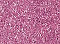
Image:ATRT-HE-Overview.jpg | AT/RT. (WC) | Image:ATRT-HE-Overview.jpg | AT/RT. (WC/marvin101) | ||
Image:ATRT-INI1.jpg | AT/RT -ve for INI1. (WC) | Image:ATRT-INI1.jpg | AT/RT -ve for INI1. (WC/marvin101) | ||
Image:Rhabdoid cells ATRT1.jpg | Rhabdoid cells in a AT/RT (WC/jensflorian) | |||
Image:ATRT-MRI.jpg | Although many of them are infratentorial, AT/RT may be found anywhere in the brain or spinal cord (WC/marvin101) | |||
</gallery> | </gallery> | ||
www: | www: | ||
Revision as of 09:55, 1 April 2015
| Atypical teratoid/rhabdoid tumour | |
|---|---|
| Diagnosis in short | |
 AT/RT. H&E stain. | |
|
| |
| LM | cellular tumour with small round cells usu. with a prominent nucleolus, rhabdoid cells (eosinophilic granular cytoplasm + eccentric nucleus), mitoses. +/-necrosis (common) |
| LM DDx | primitive neuroectodermal tumour (PNET), medulloblastoma, diffuse astrocytoma, choroid plexus carcinoma,embryonal carcinoma |
| IHC | INI1 -ve, S-100 +ve, EMA +ve, SMA +ve |
| Site | CNS - typically supratentorial |
|
| |
| Clinical history | usu. <3 years olds, occasionally adults |
| Prevalence | uncommon - esp. in adults |
| Prognosis | very poor |
Atypical teratoid/rhabdoid tumour, abbreviated AT/RT, is malignant tumour usually found supratentorially.
It may be written atypical teratoid rhabdoid tumour (abbreviated ATRT) or atypical teratoid-rhabdoid tumour (abbreviated AT-RT).
It should not be confused with the extrarenal malignant rhabdoid tumour.
General
- Usually supratentorial, occasionally in posterior fossa, case reports of spinal cord.
- Individuals usually <3 years old, uncommon in adults.[1]
- Prognosis very poor.[1]
Microscopic
Features:
- Cellular.
- Small round cells usu. with a prominent nucleolus.
- Rhabdoid cells.
- Cells with eosinophilic granular cytoplasm + eccentric nucleus.
- Mitoses.
- +/-Necrosis (common).
DDx:
- Primitive neuroectodermal tumour (PNET).
- Medulloblastoma.
- Diffuse astrocytoma.
- Choroid plexus carcinoma.
- Embryonal carcinoma.
Images
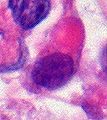
Rhabdoid tumour cell. (WC/marvin101)
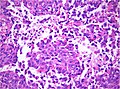
AT/RT. (WC/marvin101)
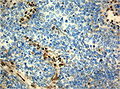
AT/RT -ve for INI1. (WC/marvin101)
Rhabdoid cells in a AT/RT (WC/jensflorian)
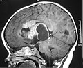
Although many of them are infratentorial, AT/RT may be found anywhere in the brain or spinal cord (WC/marvin101)


www:
IHC
- BAF-47 -ve (AKA INI1, AKA SMARCB1 - the HGNC symbol[2]) - virtually diagnostic (4/4 cases[3]).
- Endothelial cells +ve control.
- S-100 +ve (4/4 cases[3]).
- Few other brain tumours express it.
- Vimentin +ve - perinuclear condensation (4/4 cases[3]).
Others:
- GFAP +ve (focal - in tumour cells).
- EMA +ve - patchy cytoplasmic (4/4 cases[3]).
- Smooth muscle actin +ve.(4/4 cases[3]).
- Cytokeratin +ve.[citation needed]
See also
References
- ↑ 1.0 1.1 Kanoto, M.; Toyoguchi, Y.; Hosoya, T.; Kuchiki, M.; Sugai, Y. (Jan 2014). "Radiological Image Features of the Atypical Teratoid/Rhabdoid Tumor in Adults: A Systematic Review.". Clin Neuroradiol. doi:10.1007/s00062-013-0282-2. PMID 24477665.
- ↑ Online 'Mendelian Inheritance in Man' (OMIM) 601607
- ↑ 3.0 3.1 3.2 3.3 3.4 Ertan, Y.; Sezak, M.; Turhan, T.; Kantar, M.; Erşahin, Y.; Mutluer, S.; Vergin, C.; Oniz, H. et al. (Jun 2009). "Atypical teratoid/rhabdoid tumor of the central nervous system: clinicopathologic and immunohistochemical features of four cases.". Childs Nerv Syst 25 (6): 707-11. doi:10.1007/s00381-009-0811-0. PMID 19212771.