Difference between revisions of "Atypical ductal hyperplasia"
Jump to navigation
Jump to search
| Line 1: | Line 1: | ||
{{ Infobox diagnosis | |||
| Name = {{PAGENAME}} | |||
| Image = | |||
| Width = | |||
| Caption = | |||
| Synonyms = | |||
| Micro = cytologic and architectural features of low-grade DCIS (equal cell spacing, lumina round, variable architecture (classically [[cribriform]] or solid - may be micropapillary or papillary), small nuclei, small indistinct nucleoli); limited extent - either (1) two or less complete ducts, (2) <2 mm | |||
| Subtypes = | |||
| LMDDx = [[ductal carcinoma in situ]], [[invasive ductal carcinoma of the breast]] | |||
| Stains = | |||
| IHC = | |||
| EM = | |||
| Molecular = | |||
| IF = | |||
| Gross = | |||
| Grossing = | |||
| Staging = | |||
| Site = [[breast]] | |||
| Assdx = | |||
| Syndromes = | |||
| Clinicalhx = | |||
| Signs = | |||
| Symptoms = none | |||
| Prevalence = relatively common | |||
| Bloodwork = | |||
| Rads = suspicious calcifications | |||
| Endoscopy = | |||
| Prognosis = benign, increased risk of malignancy | |||
| Other = | |||
| ClinDDx = | |||
| Tx = lumpectomy when found on biopsy, follow-up if on extensional specimen | |||
}} | |||
'''Atypical ductal hyperplasia''', abbreviated '''ADH''', a benign [[breast pathology|breast lesion]] associated with an increased risk of [[malignancy]]. | '''Atypical ductal hyperplasia''', abbreviated '''ADH''', a benign [[breast pathology|breast lesion]] associated with an increased risk of [[malignancy]]. | ||
| Line 11: | Line 43: | ||
==Microscopic== | ==Microscopic== | ||
Features: | Features: | ||
*Cytologic and architectural | *Cytologic and architectural features of low-grade DCIS. | ||
**Cell spacing ~ equal. | **Cell spacing ~ equal. | ||
**Lumina round. | **Lumina round. | ||
| Line 22: | Line 54: | ||
DDx: | DDx: | ||
*Low-grade [[ | *Low-grade [[ductal carcinoma in situ]] (DCIS). | ||
*[[Florid epithelial hyperplasia of the usual type]] (FEHUT). | *[[Florid epithelial hyperplasia of the usual type]] (FEHUT). | ||
Revision as of 15:36, 28 April 2016
| Atypical ductal hyperplasia | |
|---|---|
| Diagnosis in short | |
|
| |
| LM | cytologic and architectural features of low-grade DCIS (equal cell spacing, lumina round, variable architecture (classically cribriform or solid - may be micropapillary or papillary), small nuclei, small indistinct nucleoli); limited extent - either (1) two or less complete ducts, (2) <2 mm |
| LM DDx | ductal carcinoma in situ, invasive ductal carcinoma of the breast |
| Site | breast |
|
| |
| Symptoms | none |
| Prevalence | relatively common |
| Radiology | suspicious calcifications |
| Prognosis | benign, increased risk of malignancy |
| Treatment | lumpectomy when found on biopsy, follow-up if on extensional specimen |
Atypical ductal hyperplasia, abbreviated ADH, a benign breast lesion associated with an increased risk of malignancy.
General
- Molecular studies have shown it is the same thing as low-grade DCIS; thus, some have called for abolition of the term.[1]
- ADH is considered an indication for a lumpectomy.[2]
Epidemiology:
- Relative risk of breast cancer, based on a median follow-up of 8 years, in a case control study of US registered nurses, is 3.7.[5]
Microscopic
Features:
- Cytologic and architectural features of low-grade DCIS.
- Cell spacing ~ equal.
- Lumina round.
- Architecture - classically cribriform or solid; may be micropapillary or papillary.
- Small nuclei.
- Small indistinct nucleoli.
- Limited extent (diagnostic size cutoffs) - either:[6]
- < Two complete ducts.
- < 2 mm. ‡
DDx:
- Low-grade ductal carcinoma in situ (DCIS).
- Florid epithelial hyperplasia of the usual type (FEHUT).
Notes:
- High-grade DCIS is not in the DDx of ADH.
- ‡ 3 mm is used in papillary lesions.[citation needed]
Images
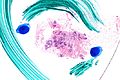
ADH. Very low mag. (WC/Nephron)
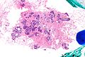
ADH - low mag. (WC/Nephron)
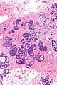
ADH - intermed. mag. (WC/Nephron)
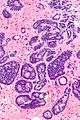
ADH - high mag. (WC/Nephron)
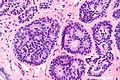
ADH - very high mag. (WC/Nephron)


IHC
- CK5 <20% +ve.
- ER +ve - diffusely.
- Heterogenous in FEHUT.
See also
References
- ↑ Ghofrani, M.; Tapia, B.; Tavassoli, FA. (Dec 2006). "Discrepancies in the diagnosis of intraductal proliferative lesions of the breast and its management implications: results of a multinational survey.". Virchows Arch 449 (6): 609-16. doi:10.1007/s00428-006-0245-y. PMID 17058097.
- ↑ Liberman L, Cohen MA, Dershaw DD, Abramson AF, Hann LE, Rosen PP (May 1995). "Atypical ductal hyperplasia diagnosed at stereotaxic core biopsy of breast lesions: an indication for surgical biopsy". AJR Am J Roentgenol 164 (5): 1111–3. PMID 7717215. http://www.ajronline.org/cgi/pmidlookup?view=long&pmid=7717215.
- ↑ Deshaies, I.; Provencher, L.; Jacob, S.; Côté, G.; Robert, J.; Desbiens, C.; Poirier, B.; Hogue, JC. et al. (Feb 2011). "Factors associated with upgrading to malignancy at surgery of atypical ductal hyperplasia diagnosed on core biopsy.". Breast 20 (1): 50-5. doi:10.1016/j.breast.2010.06.004. PMID 20619647.
- ↑ Margenthaler, JA.; Duke, D.; Monsees, BS.; Barton, PT.; Clark, C.; Dietz, JR. (Oct 2006). "Correlation between core biopsy and excisional biopsy in breast high-risk lesions.". Am J Surg 192 (4): 534-7. doi:10.1016/j.amjsurg.2006.06.003. PMID 16978969.
- ↑ London, SJ.; Connolly, JL.; Schnitt, SJ.; Colditz, GA. (Feb 1992). "A prospective study of benign breast disease and the risk of breast cancer.". JAMA 267 (7): 941-4. PMID 1734106.
- ↑ Tadrous, Paul.J. Diagnostic Criteria Handbook in Histopathology: A Surgical Pathology Vade Mecum (1st ed.). Wiley. pp. 258. ISBN 978-0470519035.