Difference between revisions of "Angiosarcoma"
(→Images) |
|||
| Line 72: | Line 72: | ||
Image:Epithelioid_angiosarcoma_-_very_high_mag.jpg | Epithelioid angiosarcoma - very high mag. (WC/Nephron) | Image:Epithelioid_angiosarcoma_-_very_high_mag.jpg | Epithelioid angiosarcoma - very high mag. (WC/Nephron) | ||
Image:Epithelioid_angiosarcoma_-_CD31_-_intermed_mag.jpg | Epithelioid angiosarcoma - CD31 - intermed. mag. (WC/Nephron) | Image:Epithelioid_angiosarcoma_-_CD31_-_intermed_mag.jpg | Epithelioid angiosarcoma - CD31 - intermed. mag. (WC/Nephron) | ||
Image:Breast Angiosarcoma Epithelioid HP2 CTR.jpg|Breast - Epithelioid angiosarcoma - high power (SKB) | |||
Image:Breast Angiosarcoma Epithelioid HP CTR.jpg|Breast - Epithelioid angiosarcoma - high power (SKB) | |||
Image:Breast Angiosarcoma Epithelioid MP CTR.jpg|Breast - Epithelioid angiosarcoma - medium power (SKB) | |||
Image:Breast Angiosarcoma HighGrade Breast HP PA.JPG|Breast - Angiosarcoma - high grade - high power (SKB) | |||
Image:Breast Angiosarcoma HighGrade Breast PA.JPG|Breast - Angiosarcoma - high grade - high power (SKB) | |||
Image:Breast Angiosarcoma HighGrade HP PA.JPG|Breast - Angiosarcoma - high grade - high power (SKB) | |||
Image:Breast Angiosarcoma HP3 SNP.jpg|Breast - Angiosarcoma - high power (SKB) | |||
Image:Breast Angiosarcoma LowGradeArea FatInvasion MP CTR.jpg|Breast - Angiosarcoma - low grade area - encircling fat - high power (SKB) | |||
Image:Breast Angiosarcoma HP SNP.jpg|Breast - Angiosarcoma - high power (SKB) | |||
Image:Breast Angiosarcoma HighGradeArea MP CTR.jpg|Breast - Angiosarcoma - high grade area - high power (SKB) | |||
Image:Breast Angiosarcoma HP PA.JPG|Breast - Angiosarcoma - high power (SKB) | |||
Image:Breast Angiosarcoma HighGrade PA.JPG|Breast - Angiosarcoma - high power (SKB) | |||
Image:Breast Angiosarcoma LP SNP.jpg|Breast - Angiosarcoma - low power (SKB) | |||
Image:Breast Angiosarcoma MP SNP.jpg|Breast - Angiosarcoma - Medium power (SKB) | |||
Image:Breast Angiosarcoma LP CTR.jpg|Breast - Angiosarcoma - low power (SKB) | |||
</gallery> | </gallery> | ||
Revision as of 11:59, 18 March 2015
| Angiosarcoma | |
|---|---|
| Diagnosis in short | |
 Epithelioid angiosarcoma. H&E stain. | |
|
| |
| LM | atypical cells - usu. spindle cells, occasionally epithelioid; vascular differentiation (abundant capillaries - "red" at low power, +/-cytoplasmic vacuolization, +/-hobnail endothelial cells) |
| LM DDx | Kaposi sarcoma, other vascular tumours |
| IHC | CD31 +ve, FLI-1 +ve, CD34 +ve, HHV-8 -ve |
| Molecular | +/-MYC amplification |
| Gross | red lesion[citation needed] |
| Site | skin, head and neck, elsewhere |
|
| |
| Syndromes | Stewart–Treves syndrome |
|
| |
| Clinical history | +/-chronic lymphedema, vinyl chloride exposure (liver angiosarcoma) |
| Prevalence | uncommon |
| Prognosis | poor |
Angiosarcoma is an uncommon malignant vascular tumour.
General
- Malignant tumour - general has a poor prognosis.[1]
Epidemiology:
- May arise secondary to chronic lymphedema related to breast carcinoma.
- Known as Stewart–Treves syndrome.[2]
- Liver angiosarcomas are associated with vinyl chloride exposure.[3]
- Cutaneous angiosarcomas are classically seen on the head and neck of whites over 60 years old.[4]
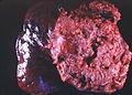
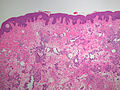
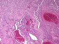
Gross
- Red/dark tan lesion.
- Typically poorly circumscribed.
Angiosarcoma. (WC/Yale Rosen)
.jpg)
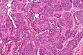
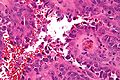
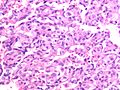
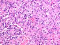
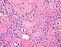
Microscopic
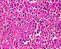
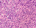
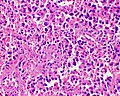
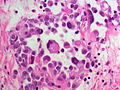
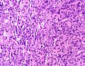
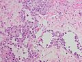
Features:
- Spindle cell lesion.
- Occasionally an epithelioid lesion.
- Very many small capillaries of irregular shape lined with:
- Pleomorphic nuclei - important.
- May have hobnail morphology.
- Usually "red" at low power - due to many RBCs - important.
- Pleomorphic nuclei - important.
- Mitoses.
- Cytoplasmic vacuoles.
- Cells trying to form lumina - embryologic.
Notes:
- Epithelioid variant (with abundant cytoplasm & sheeting architecture) may resemble melanoma or hepatocellular carcinoma.
DDx:
- Atypical vascular lesion.
- Kaposi sarcoma.
- Poorly differentiated carcinoma.
Images
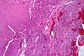
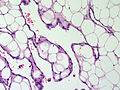
Epithelioid angiosarcoma - very low mag. (WC/Nephron)
Epithelioid angiosarcoma - intermed mag. (WC/Nephron)
Epithelioid angiosarcoma - very high mag. (WC/Nephron)
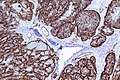
Epithelioid angiosarcoma - CD31 - intermed. mag. (WC/Nephron)
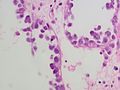
Breast - Epithelioid angiosarcoma - high power (SKB)
Breast - Epithelioid angiosarcoma - high power (SKB)
Breast - Epithelioid angiosarcoma - medium power (SKB)
Breast - Angiosarcoma - high grade - high power (SKB)
Breast - Angiosarcoma - high grade - high power (SKB)
Breast - Angiosarcoma - high grade - high power (SKB)
Breast - Angiosarcoma - high power (SKB)
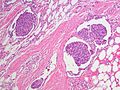
Breast - Angiosarcoma - low grade area - encircling fat - high power (SKB)
Breast - Angiosarcoma - high power (SKB)
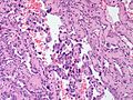
Breast - Angiosarcoma - high grade area - high power (SKB)
Breast - Angiosarcoma - high power (SKB)
Breast - Angiosarcoma - high power (SKB)
Breast - Angiosarcoma - low power (SKB)
Breast - Angiosarcoma - Medium power (SKB)
Breast - Angiosarcoma - low power (SKB)


IHC
Molecular
See also
References
- ↑ Young RJ, Brown NJ, Reed MW, Hughes D, Woll PJ (May 2010). "Angiosarcoma". Lancet Oncol. doi:10.1016/S1470-2045(10)70023-1. PMID 20537949.
- ↑ Pincus, LB.; Fox, LP. (Aug 2008). "Images in clinical medicine. The Stewart-Treves syndrome.". N Engl J Med 359 (9): 950. doi:10.1056/NEJMicm071344. PMID 18753651. http://www.nejm.org/doi/full/10.1056/NEJMicm071344.
- ↑ Mitchell, Richard; Kumar, Vinay; Fausto, Nelson; Abbas, Abul K.; Aster, Jon (2011). Pocket Companion to Robbins & Cotran Pathologic Basis of Disease (8th ed.). Elsevier Saunders. pp. 212. ISBN 978-1416054542.
- ↑ Albores-Saavedra, J.; Schwartz, AM.; Henson, DE.; Kostun, L.; Hart, A.; Angeles-Albores, D.; Chablé-Montero, F. (Apr 2011). "Cutaneous angiosarcoma. Analysis of 434 cases from the Surveillance, Epidemiology, and End Results Program, 1973-2007.". Ann Diagn Pathol 15 (2): 93-7. doi:10.1016/j.anndiagpath.2010.07.012. PMID 21190880.
- ↑ Rossi, S.; Orvieto, E.; Furlanetto, A.; Laurino, L.; Ninfo, V.; Dei Tos, AP. (May 2004). "Utility of the immunohistochemical detection of FLI-1 expression in round cell and vascular neoplasm using a monoclonal antibody.". Mod Pathol 17 (5): 547-52. doi:10.1038/modpathol.3800065. PMID 15001993.
- ↑ Kahn, HJ.; Bailey, D.; Marks, A. (Apr 2002). "Monoclonal antibody D2-40, a new marker of lymphatic endothelium, reacts with Kaposi's sarcoma and a subset of angiosarcomas.". Mod Pathol 15 (4): 434-40. doi:10.1038/modpathol.3880543. PMID 11950918.
- ↑ Kurisetty, V.; Bryan, BA. (Apr 2013). "Aberrations in Angiogenic Signaling and MYC Amplifications are Distinguishing Features of Angiosarcoma.". Angiol Open Access 1. doi:10.4172/2329-9495.1000102. PMID 25374893.
- ↑ Styring, E.; Seinen, J.; Dominguez-Valentin, M.; Domanski, HA.; Jönsson, M.; von Steyern, FV.; Hoekstra, HJ.; Suurmeijer, AJ. et al. (Jul 2014). "Key roles for MYC, KIT and RET signaling in secondary angiosarcomas.". Br J Cancer 111 (2): 407-12. doi:10.1038/bjc.2014.359. PMID 24983371.