Warthin tumour
Jump to navigation
Jump to search
Warthin tumour is a relative common benign tumour of the parotid gland. It is also known as papillary cystadenoma lymphomatosum.
| Warthin tumour | |
|---|---|
| Diagnosis in short | |
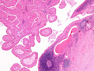
 Warthin tumour. H&E stain. | |
|
| |
| LM | papillae with a two rows of pink (eosinophilic) epithelial cells (with cuboidal basal cells and columnar luminal cells), fibrous capsule, cystic space filled with debris, lymphoid stroma |
| LM DDx | lymphoepithelial cyst |
| Gross | classically cystic, motor oil-like fluid |
| Site | salivary gland - parotid gland only |
|
| |
| Clinical history | strong association with smoking |
| Signs | mass lesion |
| Prevalence | common benign salivary gland lesion |
| Prognosis | good, benign |
| Clin. DDx | other salivary gland tumours |
General
- Benign.
Epidemiology:
- May be multicentric ~ 15% of the time.
- Bilateral ~6% of the time.[1]
- Classically: male > female - changing with more women smokers.
- Smokers - almost 80% of patients is a series of 70 cases.[1]
- Old - usually 60s,[2] very rarely < 40 years old.
Notes:
- No malignant transformation.
- Not in submandibular gland.
- Not in sublingual gland.
- Not in children.
Gross
Features:
- Motor oil-like fluid.[3]
- Cystic component larger in larger lesions.
- Small lesions may be solid.
Image:
Microscopic
Features:
- Papillae (nipple-shaped structures) with a two rows of pink (eosinophilic) epithelial cells (with cuboidal basal cells and columnar luminal cells) - key feature.
- Fibrous capsule - pink & homogenous on H&E stain.
- Cystic space filled with debris in situ (not necrosis).
- Lymphoid stroma.
Notes:
- +/-Squamous differentiation.
- +/-Goblet cell differentiation.
DDx:
- Lymphoepithelial cyst.
- Cyst within a lymph node.
- Lymphoma associated with Warthin tumour - case reports.[4][5]
Images
Case 1
Case 2
Sign out
Parotid Gland, Left, Excision:
- Warthin's tumour (papillary cystadenoma lymphomatosum).
- NEGATIVE for malignancy.
Block letters
PAROTID GLAND, RIGHT, EXCISION:
- WARTHIN TUMOUR.
Micro
The sections show a cystic tumour with lymphoid tissue associated with benign salivary gland tissue. The lymphoid tissue is composed of small cells and forms morphologically unremarkable follicles. The cyst-lining epithelium has a bilayered appearance and is composed of cells with abundant eosinophilic cytoplasm and nucleoli. The tumour focally extends to the edge of the tissue (ink present on tumour).
See also
References
- ↑ 1.0 1.1 Chedid, HM.; Rapoport, A.; Aikawa, KF.; Menezes, Ados S.; Curioni, OA.. "Warthin's tumor of the parotid gland: study of 70 cases.". Rev Col Bras Cir 38 (2): 90-4. PMID 21710045.
- ↑ Dăguci, L.; Simionescu, C.; Stepan, A.; Munteanu, C.; Dăguci, C.; Bătăiosu, M. (2011). "Warthin tumor--morphological study of the stromal compartment.". Rom J Morphol Embryol 52 (4): 1319-23. PMID 22203940.
- ↑ Hunt, JL. (Jan 2006). "Warthin tumors do not have microsatellite instability and express normal DNA mismatch repair proteins.". Arch Pathol Lab Med 130 (1): 52-6. doi:10.1043/1543-2165(2006)130[52:WTDNHM]2.0.CO;2. PMID 16390238.
- ↑ Alnoor, F.; Gandhi, JS.; Stein, MK.; Gradowski, JF. (Jun 2019). "Follicular Lymphoma Diagnosed in Warthin Tumor: A Case Report and Review of the Literature.". Head Neck Pathol. doi:10.1007/s12105-019-01045-x. PMID 31183747.
- ↑ Jawad, H.; McCarthy, P.; O'Leary, G.; Heffron, CC. (May 2018). "Presentation of Chronic Lymphocytic Leukemia/Small Lymphocytic Lymphoma in a Warthin Tumor: Case Report and Literature Review.". Int J Surg Pathol 26 (3): 256-260. doi:10.1177/1066896917734371. PMID 28978260.